1B9C

Green Fluorescent Protein Mutant F99S, M153T and V163A


Experimental Data Snapshot

  • Method: X-RAY DIFFRACTION
  • Resolution: 2.40 Å
  • R-Value Free: 0.280 
  • R-Value Work: 0.204 
  • R-Value Observed: 0.204 

wwPDB Validation   3D Report Full Report


This is version 1.5 of the entry. See complete history


Literature

Crystal structure and refolding properties of the mutant F99S/M153T/V163A of the green fluorescent protein.

Battistutta, R.Negro, A.Zanotti, G.

(2000) Proteins 41: 429-437

  • DOI: https://doi.org/10.1002/1097-0134(20001201)41:4<429::aid-prot10>3.0.co;2-d
  • Primary Citation of Related Structures:  
    1B9C

  • PubMed Abstract: 

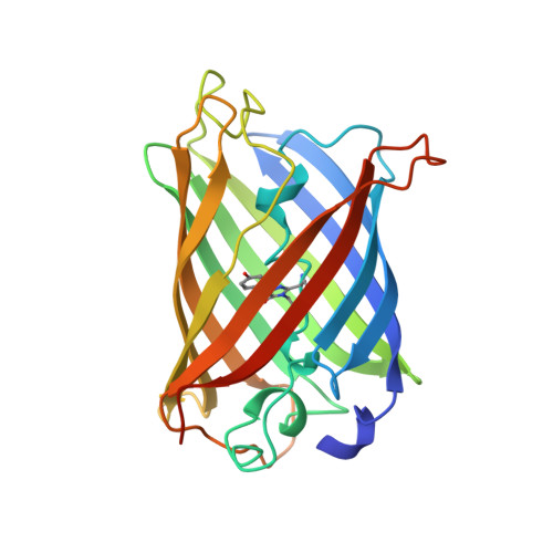
    The mutant F99S/M153T/V163A of the Green Fluorescent Protein (c3-GFP) has spectral characteristics similar to the wild-type GFP, but it is 42-fold more fluorescent in vivo. Here, we report the crystal structure and the refolding properties of c3-GFP and compare them with those of the less fluorescent wt-GFP and S65T mutant. The topology and the overall structure of c3-GFP is similar to the wild-type GFP. The three mutated residues, Ser99, Thr153, and Ala163, lie on the surface of the protein in three different beta-strands. The side chains of Ser99 and Thr153 are exposed to the solvent, whereas that of Ala163 points toward the interior of the protein. No significant deviation from the structure of the wild-type molecule is found around these positions, and there is not clear evidence of any distortion in the position of the chromophore or of the surrounding residues induced by the mutated amino acids. In vitro refolding experiments on urea-denatured c3-GFP reveal a renaturation behavior similar to that of the S65T molecule, with kinetic constants of the same order of magnitude. We conclude that the higher fluorescence activity of c3-GFP can be attributed neither to particular structural features nor to a faster folding process, as previously proposed.


  • Organizational Affiliation

    Dipartimento di Chimica Organica and Centro Studi sui Biopolimeri, Università di Padova, Padova, Italy. roberto@chor.unipd.it


Macromolecules
Find similar proteins by:  (by identity cutoff)  |  3D Structure
Entity ID: 1
MoleculeChains Sequence LengthOrganismDetailsImage
PROTEIN (GREEN FLUORESCENT PROTEIN)
A, B, C, D
236Aequorea victoriaMutation(s): 5 
UniProt
Find proteins for P42212 (Aequorea victoria)
Explore P42212 
Go to UniProtKB:  P42212
Entity Groups  
Sequence Clusters30% Identity50% Identity70% Identity90% Identity95% Identity100% Identity
UniProt GroupP42212
Sequence Annotations
Expand
  • Reference Sequence
Small Molecules
Modified Residues  1 Unique
IDChains TypeFormula2D DiagramParent
CRO
Query on CRO
A, B, C, D
L-PEPTIDE LINKINGC15 H17 N3 O5THR, TYR, GLY
Experimental Data & Validation

Experimental Data

  • Method: X-RAY DIFFRACTION
  • Resolution: 2.40 Å
  • R-Value Free: 0.280 
  • R-Value Work: 0.204 
  • R-Value Observed: 0.204 
  • Space Group: P 21 21 21
Unit Cell:
Length ( Å )Angle ( ˚ )
a = 83.48α = 90
b = 85.37β = 90
c = 140.06γ = 90
Software Package:
Software NamePurpose
AMoREphasing
X-PLORrefinement
MOSFLMdata reduction
CCP4data scaling

Structure Validation

View Full Validation Report


Entry History 

Deposition Data

Revision History  (Full details and data files)

  • Version 1.0: 2000-11-17
    Type: Initial release
  • Version 1.1: 2008-04-26
    Changes: Version format compliance
  • Version 1.2: 2011-07-13
    Changes: Version format compliance
  • Version 1.3: 2018-03-14
    Changes: Database references
  • Version 1.4: 2023-08-09
    Changes: Data collection, Database references, Derived calculations, Refinement description
  • Version 1.5: 2023-11-15
    Changes: Data collection