1BM1

CRYSTAL STRUCTURE OF BACTERIORHODOPSIN IN THE LIGHT-ADAPTED STATE


Experimental Data Snapshot

  • Method: X-RAY DIFFRACTION
  • Resolution: 3.50 Å
  • R-Value Free: 0.311 
  • R-Value Work: 0.260 
  • R-Value Observed: 0.260 

wwPDB Validation   3D Report Full Report


This is version 2.0 of the entry. See complete history


Literature

Specific lipid-protein interactions in a novel honeycomb lattice structure of bacteriorhodopsin.

Sato, H.Takeda, K.Tani, K.Hino, T.Okada, T.Nakasako, M.Kamiya, N.Kouyama, T.

(1999) Acta Crystallogr D Biol Crystallogr 55: 1251-1256

  • DOI: https://doi.org/10.1107/s090744499900503x
  • Primary Citation of Related Structures:  
    1BM1

  • PubMed Abstract: 

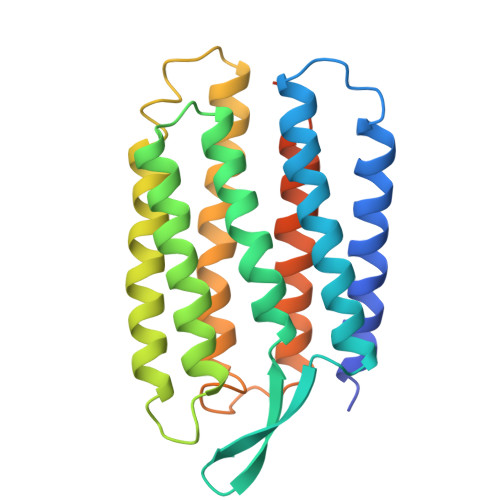
    In the purple membrane of Halobacterium salinarium, bacteriorhodopsin trimers are arranged in a hexagonal lattice. When purple membrane sheets are incubated at high temperature with neutral detergent, membrane vesicularization takes place, yielding inside-out vesicles with a diameter of 50 nm. The vesicular structure becomes unstable at low temperature, where successive fusion of the vesicles yields a crystal which is composed of stacked planar membranes. X-ray crystallographic analysis reveals that the bacteriorhodopsin trimers are arranged in a honeycomb lattice in each membrane layer and that neighbouring membranes orient in opposite directions. The native structure of the trimeric unit is preserved in the honeycomb lattice, irrespective of alterations in the in-plane orientation of the trimer. One phospholipid tightly bound to a crevice between monomers in the trimeric unit is suggested to act as a glue in the formation of the trimer.


  • Organizational Affiliation

    Department of Physics, Graduate School of Science, Nagoya University, Nagoya 464-8602, Japan.


Macromolecules
Find similar proteins by:  (by identity cutoff)  |  3D Structure
Entity ID: 1
MoleculeChains Sequence LengthOrganismDetailsImage
BACTERIORHODOPSIN248Halobacterium salinarumMutation(s): 0 
Membrane Entity: Yes 
UniProt
Find proteins for P02945 (Halobacterium salinarum (strain ATCC 700922 / JCM 11081 / NRC-1))
Explore P02945 
Go to UniProtKB:  P02945
Entity Groups  
Sequence Clusters30% Identity50% Identity70% Identity90% Identity95% Identity100% Identity
UniProt GroupP02945
Sequence Annotations
Expand
  • Reference Sequence
Small Molecules
Ligands 2 Unique
IDChains Name / Formula / InChI Key2D Diagram3D Interactions
DPG
Query on DPG

Download Ideal Coordinates CCD File 
C [auth A]PHOSPHORIC ACID 2,3-BIS-(3,7,11,15-TETRAMETHYL-HEXADECYLOXY)-PROPYL ESTER 2-HYDROXO-3-PHOSPHONOOXY-PROPYL ESTER
C46 H96 O11 P2
TZXJQSKPTCRGCA-QVBLKRSVSA-N
RET
Query on RET

Download Ideal Coordinates CCD File 
B [auth A]RETINAL
C20 H28 O
NCYCYZXNIZJOKI-OVSJKPMPSA-N
Modified Residues  1 Unique
IDChains TypeFormula2D DiagramParent
PCA
Query on PCA
A
L-PEPTIDE LINKINGC5 H7 N O3GLN
Experimental Data & Validation

Experimental Data

  • Method: X-RAY DIFFRACTION
  • Resolution: 3.50 Å
  • R-Value Free: 0.311 
  • R-Value Work: 0.260 
  • R-Value Observed: 0.260 
  • Space Group: P 6 2 2
Unit Cell:
Length ( Å )Angle ( ˚ )
a = 104.7α = 90
b = 104.7β = 90
c = 114.1γ = 120
Software Package:
Software NamePurpose
X-PLORmodel building
X-PLORrefinement
WEISdata reduction
CCP4data scaling
X-PLORphasing

Structure Validation

View Full Validation Report


Entry History 

Deposition Data

Revision History  (Full details and data files)

  • Version 1.0: 1999-04-27
    Type: Initial release
  • Version 1.1: 2008-03-24
    Changes: Version format compliance
  • Version 1.2: 2011-07-13
    Changes: Derived calculations, Version format compliance
  • Version 2.0: 2023-08-02
    Changes: Database references, Derived calculations, Other, Polymer sequence, Refinement description