1C4F

GREEN FLUORESCENT PROTEIN S65T AT PH 4.6


Experimental Data Snapshot

  • Method: X-RAY DIFFRACTION
  • Resolution: 2.25 Å
  • R-Value Work: 0.193 

wwPDB Validation   3D Report Full Report


This is version 1.6 of the entry. See complete history


Literature

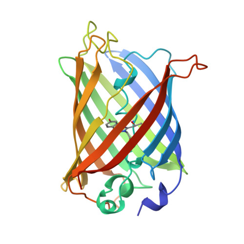
Structural and spectral response of green fluorescent protein variants to changes in pH.

Elsliger, M.A.Wachter, R.M.Hanson, G.T.Kallio, K.Remington, S.J.

(1999) Biochemistry 38: 5296-5301

  • DOI: https://doi.org/10.1021/bi9902182
  • Primary Citation of Related Structures:  
    1C4F, 1EMG

  • PubMed Abstract: 

    The green fluorescent protein (GFP) from the jellyfish Aequorea victoria has become a useful tool in molecular and cell biology. Recently, it has been found that the fluorescence spectra of most mutants of GFP respond rapidly and reversibly to pH variations, making them useful as probes of intracellular pH. To explore the structural basis for the titration behavior of the popular GFP S65T variant, we determined high-resolution crystal structures at pH 8.0 and 4.6. The structures revealed changes in the hydrogen bond pattern with the chromophore, suggesting that the pH sensitivity derives from protonation of the chromophore phenolate. Mutations were designed in yellow fluorescent protein (S65G/V68L/S72A/T203Y) to change the solvent accessibility (H148G) and to modify polar groups (H148Q, E222Q) near the chromophore. pH titrations of these variants indicate that the chromophore pKa can be modulated over a broad range from 6 to 8, allowing for pH determination from pH 5 to pH 9. Finally, mutagenesis was used to raise the pKa from 6.0 (S65T) to 7.8 (S65T/H148D). Unlike other variants, S65T/H148D exhibits two pH-dependent excitation peaks for green fluorescence with a clean isosbestic point. This raises the interesting possibility of using fluorescence at this isosbestic point as an internal reference. Practical real time in vivo applications in cell and developmental biology are proposed.


  • Organizational Affiliation

    Institute of Molecular Biology, Department of Physics, University of Oregon, Eugene 97403, USA.


Macromolecules
Find similar proteins by:  (by identity cutoff)  |  3D Structure
Entity ID: 1
MoleculeChains Sequence LengthOrganismDetailsImage
GREEN FLUORESCENT PROTEIN236Aequorea victoriaMutation(s): 2 
UniProt
Find proteins for P42212 (Aequorea victoria)
Explore P42212 
Go to UniProtKB:  P42212
Entity Groups  
Sequence Clusters30% Identity50% Identity70% Identity90% Identity95% Identity100% Identity
UniProt GroupP42212
Sequence Annotations
Expand
  • Reference Sequence
Small Molecules
Modified Residues  1 Unique
IDChains TypeFormula2D DiagramParent
CRO
Query on CRO
A
L-PEPTIDE LINKINGC15 H17 N3 O5THR, TYR, GLY
Experimental Data & Validation

Experimental Data

  • Method: X-RAY DIFFRACTION
  • Resolution: 2.25 Å
  • R-Value Work: 0.193 
  • Space Group: P 21 21 21
Unit Cell:
Length ( Å )Angle ( ˚ )
a = 51.934α = 90
b = 62.898β = 90
c = 69.172γ = 90
Software Package:
Software NamePurpose
CCP4model building
PROTEINmodel building
TNTrefinement
DENZOdata reduction
SCALEPACKdata scaling
CCP4phasing
PROTEINphasing

Structure Validation

View Full Validation Report


Entry History 

Deposition Data

Revision History  (Full details and data files)

  • Version 1.0: 1999-08-31
    Type: Initial release
  • Version 1.1: 2007-10-16
    Changes: Version format compliance
  • Version 1.2: 2011-07-13
    Changes: Version format compliance
  • Version 1.3: 2017-10-04
    Changes: Advisory
  • Version 1.4: 2021-11-03
    Changes: Advisory, Database references, Derived calculations
  • Version 1.5: 2023-08-09
    Changes: Data collection, Refinement description
  • Version 1.6: 2023-11-15
    Changes: Data collection