1GWG

Tri-iodide derivative of apoferritin


Experimental Data Snapshot

  • Method: X-RAY DIFFRACTION
  • Resolution: 2.01 Å
  • R-Value Free: 0.208 
  • R-Value Work: 0.167 
  • R-Value Observed: 0.169 

wwPDB Validation   3D Report Full Report


This is version 1.3 of the entry. See complete history


Literature

Triiodide Derivatization and Combinatorial Counter-Ion Replacement: Two Methods for Enhancing Phasing Signal Using Laboratory Cu Kalpha X-Ray Equipment

Evans, G.Bricogne, G.

(2002) Acta Crystallogr D Biol Crystallogr 58: 976

  • DOI: https://doi.org/10.1107/s0907444902005486
  • Primary Citation of Related Structures:  
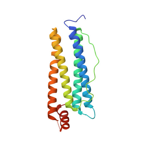
    1GW9, 1GWA, 1GWD, 1GWG

  • PubMed Abstract: 

    A series of experiments performed at Cu Kalpha wavelength on in-house X-ray equipment are presented which investigate two possibilities for enhancing the experimental phasing signal by means of (i) triiodide/iodide soaks using KI/I(2) and (ii) combinations of counter-ions introduced using the short cryosoak method. Triiodide-derivative crystal structures for five test proteins have been refined and reveal that iodine can bind as polyiodide and single iodide ions through hydrophobic and hydrogen-bonding interactions both at the molecular surface and in intramolecular and intermolecular cavities. In three cases, the structures could be automatically determined with autoSHARP using in-house SAD and SIRAS data. The investigation of combinatorial counter-ion replacement using multiple salts with Na(+) and Cs(+) as cations and I(-) and Cl(-) as anions reveals that, for the case of hen egg-white lysozyme, significant improvement in phasing signal is obtained by the combined use of salts compared with SIRAS methods using native and single short-soak derivative data sets.


  • Organizational Affiliation

    Global Phasing Limited, Cambridge, England.


Macromolecules
Find similar proteins by:  (by identity cutoff)  |  3D Structure
Entity ID: 1
MoleculeChains Sequence LengthOrganismDetailsImage
FERRITIN LIGHT CHAIN174Equus caballusMutation(s): 0 
UniProt
Find proteins for P02791 (Equus caballus)
Explore P02791 
Go to UniProtKB:  P02791
Entity Groups  
Sequence Clusters30% Identity50% Identity70% Identity90% Identity95% Identity100% Identity
UniProt GroupP02791
Sequence Annotations
Expand
  • Reference Sequence
Small Molecules
Ligands 2 Unique
IDChains Name / Formula / InChI Key2D Diagram3D Interactions
IOD
Query on IOD

Download Ideal Coordinates CCD File 
B [auth A]
C [auth A]
D [auth A]
E [auth A]
F [auth A]
B [auth A],
C [auth A],
D [auth A],
E [auth A],
F [auth A],
G [auth A],
J [auth A],
K [auth A],
L [auth A]
IODIDE ION
I
XMBWDFGMSWQBCA-UHFFFAOYSA-M
CD
Query on CD

Download Ideal Coordinates CCD File 
H [auth A],
I [auth A]
CADMIUM ION
Cd
WLZRMCYVCSSEQC-UHFFFAOYSA-N
Experimental Data & Validation

Experimental Data

  • Method: X-RAY DIFFRACTION
  • Resolution: 2.01 Å
  • R-Value Free: 0.208 
  • R-Value Work: 0.167 
  • R-Value Observed: 0.169 
  • Space Group: F 4 3 2
Unit Cell:
Length ( Å )Angle ( ˚ )
a = 181.19α = 90
b = 181.19β = 90
c = 181.19γ = 90
Software Package:
Software NamePurpose
BUSTER-TNTrefinement
MOSFLMdata reduction
SCALAdata scaling

Structure Validation

View Full Validation Report


Entry History 

Deposition Data

Revision History  (Full details and data files)

  • Version 1.0: 2002-06-06
    Type: Initial release
  • Version 1.1: 2011-05-08
    Changes: Version format compliance
  • Version 1.2: 2011-07-13
    Changes: Version format compliance
  • Version 1.3: 2017-06-28
    Changes: Data collection