Engineering the zinc binding site of human carbonic anhydrase II: structure of the His-94-->Cys apoenzyme in a new crystalline form.
Alexander, R.S., Kiefer, L.L., Fierke, C.A., Christianson, D.W.(1993) Biochemistry 32: 1510-1518
- PubMed: 8431430
- DOI: https://doi.org/10.1021/bi00057a015
- Primary Citation of Related Structures:
1HVA - PubMed Abstract:
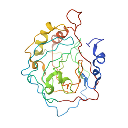
The structure of the His-94-->Cys variant of human carbonic anhydrase II (CAII) has been determined by X-ray crystallographic methods to a resolution of 2.3 A with a final crystallographic R factor of 0.155. This variant of CAII crystallizes in orthorhombic space group P2(1)2(1)2(1) which is the first example of a new crystal form for this important zinc hydrase (the wild-type enzyme crystallizes in monoclinic space group P21 under similar crystallization conditions). Although the overall structure of the enzyme in the orthorhombic crystal form is similar to that of the wild-type protein in the monoclinic crystal form, the rms deviation of C alpha atoms between the two structures is 0.5 A. Larger structural deviations occur in regions of the protein molecule involved in crystal lattice contacts, and significant structural changes are found in the polypeptide strand containing Cys-94. Surprisingly, no electron density corresponding to a zinc ion is found in the active site of crystalline His-94-->Cys CAII, even though the stoichiometry of zinc binding to this variant in solution is confirmed by atomic absorption spectroscopy. However, the KD for zinc dissociation from the variant is increased 10(4)-fold compared with wild-type enzyme; furthermore, under the crystallization conditions of high ionic strength (1.75-2.5 M ammonium sulfate), the observed KD is increased further, which leads to zinc dissociation. Spectroscopic analysis of Co(2+)-substituted His-94-->Cys CAII indicates that the metal binds in a tetrahedral geometry with a new thiolate bond.(ABSTRACT TRUNCATED AT 250 WORDS)
Organizational Affiliation:
Department of Chemistry, University of Pennsylvania, Philadelphia 19104-6323.