1IZR

F46A mutant of bovine pancreatic ribonuclease A


Experimental Data Snapshot

  • Method: X-RAY DIFFRACTION
  • Resolution: 1.50 Å
  • R-Value Free: 0.226 
  • R-Value Work: 0.198 
  • R-Value Observed: 0.198 

wwPDB Validation   3D Report Full Report


This is version 1.4 of the entry. See complete history


Literature

Minimization of cavity size ensures protein stability and folding: structures of Phe46-replaced bovine pancreatic RNase A

Kadonosono, T.Chatani, E.Hayashi, R.Moriyama, H.Ueki, T.

(2003) Biochemistry 42: 10651-10658

  • DOI: https://doi.org/10.1021/bi034499w
  • Primary Citation of Related Structures:  
    1IZP, 1IZQ, 1IZR

  • PubMed Abstract: 

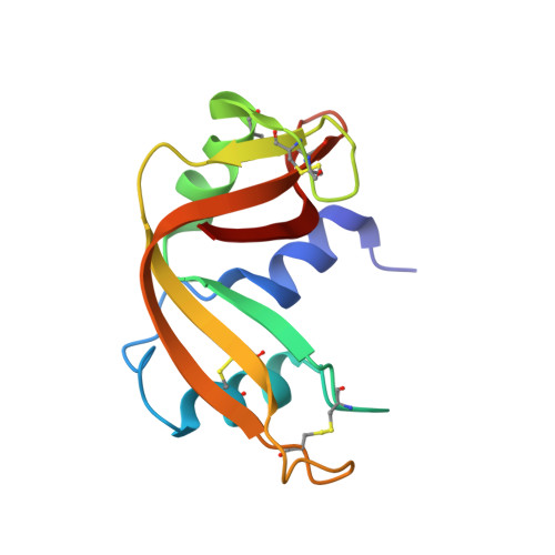
    The Phe46 residue, located in the hydrophobic core of RNase A, was replaced with other hydrophobic residues, leucine, valine, or alanine, and their X-ray crystallographic structures were determined up to 1.50-1.80 A resolution in an attempt to examine the relationship between structural changes and conformational stability or folding kinetics. The backbone structure of F46L, F46V, and F46A was indistinguishable from that of the wild-type enzyme, retaining the correct active site structure. However, one water molecule was included in the hydrophobic core of F46A, forming two hydrogen bonds with the backbone peptide chain. The side chain of Met29 in F46V and F46A adopted two different conformations in an equal occupancy. A trapped water molecule and two conformations of Met29 represent changes that minimize the cavity volume. Nevertheless, the replacement of Phe46 with the above residues resulted in a marked decrease in both thermal stability and folding reaction. Thus, Phe46 ensures the thermal stability and the rapid and correct folding of RNase A by the role it plays in forming a highly packed, hydrophobic core.


  • Organizational Affiliation

    Division of Applied Life Sciences, Graduate School of Agriculture, Kyoto University, Sakyo, Kyoto 606-8502, Japan.


Macromolecules
Find similar proteins by:  (by identity cutoff)  |  3D Structure
Entity ID: 1
MoleculeChains Sequence LengthOrganismDetailsImage
RIBONUCLEASE A124Bos taurusMutation(s): 1 
EC: 3.1.27.5
UniProt
Find proteins for P61823 (Bos taurus)
Explore P61823 
Go to UniProtKB:  P61823
Entity Groups  
Sequence Clusters30% Identity50% Identity70% Identity90% Identity95% Identity100% Identity
UniProt GroupP61823
Sequence Annotations
Expand
  • Reference Sequence
Experimental Data & Validation

Experimental Data

  • Method: X-RAY DIFFRACTION
  • Resolution: 1.50 Å
  • R-Value Free: 0.226 
  • R-Value Work: 0.198 
  • R-Value Observed: 0.198 
  • Space Group: P 32 2 1
Unit Cell:
Length ( Å )Angle ( ˚ )
a = 64.241α = 90
b = 64.241β = 90
c = 63.057γ = 120
Software Package:
Software NamePurpose
DENZOdata reduction
SCALEPACKdata scaling
CNSrefinement
CNSphasing

Structure Validation

View Full Validation Report


Entry History 

Deposition Data

Revision History  (Full details and data files)

  • Version 1.0: 2003-11-25
    Type: Initial release
  • Version 1.1: 2008-04-27
    Changes: Version format compliance
  • Version 1.2: 2011-07-13
    Changes: Version format compliance
  • Version 1.3: 2021-11-10
    Changes: Database references
  • Version 1.4: 2023-10-25
    Changes: Data collection, Refinement description