Structural evidence for a dominant role of nonpolar interactions in the binding of a transport/chemosensory receptor to its highly polar ligands.
Duan, X., Quiocho, F.A.(2002) Biochemistry 41: 706-712
- PubMed: 11790091
- DOI: https://doi.org/10.1021/bi015784n
- Primary Citation of Related Structures:
1JW4 - PubMed Abstract:
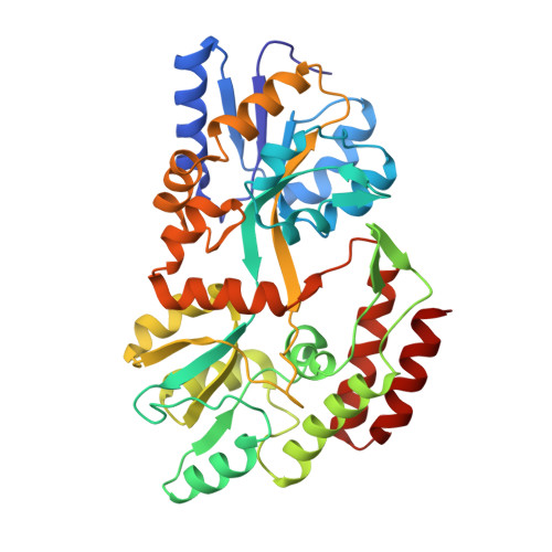
The receptor, a maltose/maltooligosaccharide-binding protein, has been found to be an excellent system for the study of molecular recognition because its polar and nonpolar binding functions are segregated into two globular domains. The X-ray structures of the "closed" and "open" forms of the protein complexed with maltose and maltotetraitol have been determined. These sugars have approximately 3 times more accessible polar surface (from OH groups) than nonpolar surface (from small clusters of sugar ring CH bonds). In the closed structures, the oligosaccharides are buried in the groove between the two domains of the protein and bound by extensive hydrogen bonding interactions of the OH groups with the polar residues confined mostly in one domain and by nonpolar interactions of the CH clusters with four aromatic residues lodged in the other domain. Substantial contacts between the sugar hydroxyls and aromatic residues are also formed. In the open structures, the oligosaccharides are bound almost exclusively in the domain rich in aromatic residues. This finding, along with the analysis of buried surface area due to complex formations in the open and closed structures, supports a major role for nonpolar interactions in initial ligand binding even when the ligands have significantly greater potential for highly specific polar interactions.
Organizational Affiliation:
Howard Hughes Medical Institute and Department of Biochemistry and Molecular Biology, Baylor College of Medicine, Houston, Texas 77030, USA.