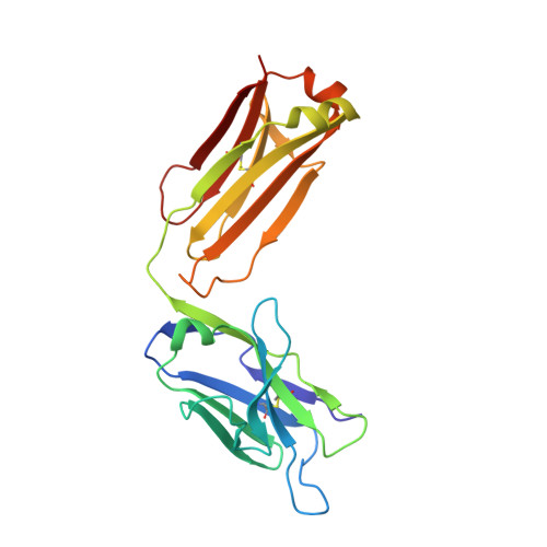
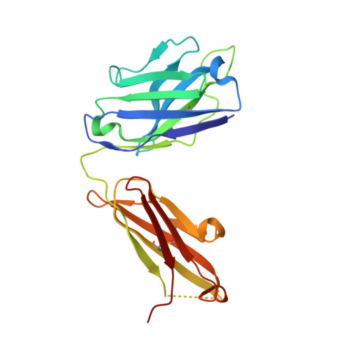
1KFA

Crystal structure of Fab fragment complexed with gibberellin A4


Experimental Data Snapshot

  • Method: X-RAY DIFFRACTION
  • Resolution: 2.80 Å
  • R-Value Free: 0.313 
  • R-Value Work: 0.235 
  • R-Value Observed: 0.261 

wwPDB Validation   3D Report Full Report


This is version 1.4 of the entry. See complete history


Literature

Crystal structure of the liganded anti-gibberellin A(4) antibody 4-B8(8)/E9 Fab fragment.

Murata, T.Fushinobu, S.Nakajima, M.Asami, O.Sassa, T.Wakagi, T.Yamaguchi, I.

(2002) Biochem Biophys Res Commun 293: 489-496

  • DOI: https://doi.org/10.1016/S0006-291X(02)00225-5
  • Primary Citation of Related Structures:  
    1KFA

  • PubMed Abstract: 

    Gibberellins, a class of plant hormones, consist of more than 120 members. Only a few of them are recognized by a receptor that remains unknown. The haptenic mouse monoclonal antibody, 4-B8(8)/E9, was generated against gibberellin A(4) (GA(4)) to recognize biologically active GA selectivity, and we attempted to confirm the binding properties between the antibody and GA(4). We carried out an X-ray crystallographic analysis of the 4-B8(8)/E9 Fab fragment complexed with GA(4) at a 2.8 A resolution by using the molecular replacement method. The crystal structure of the Fab fragment showed the typical immunoglobulin fold of the beta-barrel structure which is the common motif of all antibodies. A small hapten-combining site was made up of three heavy chain CDR loops. On the other hand, CDRs of the light chain did not interact directly with GA(4). The C/D rings of the GA(4) molecule were in van der Waals contact mainly with the aromatic side chain of Tyr100AH and Phe100BH of CDR-H3. The 3 beta-hydroxyl and 6 beta-carboxyl groups were, respectively, hydrogen-bonded to the main chain of Ala33H and to the Thr53H heavy chain.


  • Organizational Affiliation

    Department of Applied Biological Chemistry, Division of Agriculture and Agricultural Life Sciences, The University of Tokyo, 1-1-1 Yayoi, Bunkyo-ku, Tokyo 113-8657, Japan. tks@pgr1.ch.a.u-tokyo.ac.jp


Macromolecules
Find similar proteins by:  (by identity cutoff)  |  3D Structure
Entity ID: 1
MoleculeChains Sequence LengthOrganismDetailsImage
monoclonal antibody light chainA [auth L],
C [auth M]
217Mus musculusMutation(s): 0 
Entity Groups  
Sequence Clusters30% Identity50% Identity70% Identity90% Identity95% Identity100% Identity
Sequence Annotations
Expand
  • Reference Sequence
Find similar proteins by:  (by identity cutoff)  |  3D Structure
Entity ID: 2
MoleculeChains Sequence LengthOrganismDetailsImage
monoclonal antibody heavy chainB [auth H],
D [auth I]
221Mus musculusMutation(s): 0 
Entity Groups  
Sequence Clusters30% Identity50% Identity70% Identity90% Identity95% Identity100% Identity
Sequence Annotations
Expand
  • Reference Sequence
Experimental Data & Validation

Experimental Data

  • Method: X-RAY DIFFRACTION
  • Resolution: 2.80 Å
  • R-Value Free: 0.313 
  • R-Value Work: 0.235 
  • R-Value Observed: 0.261 
  • Space Group: C 2 2 21
Unit Cell:
Length ( Å )Angle ( ˚ )
a = 118.076α = 90
b = 150.146β = 90
c = 106.87γ = 90
Software Package:
Software NamePurpose
MOSFLMdata reduction
SCALAdata scaling
CNSrefinement
CCP4data scaling
CNSphasing

Structure Validation

View Full Validation Report


Entry History 

Deposition Data

Revision History  (Full details and data files)

  • Version 1.0: 2002-09-11
    Type: Initial release
  • Version 1.1: 2008-04-27
    Changes: Version format compliance
  • Version 1.2: 2011-07-13
    Changes: Version format compliance
  • Version 1.3: 2013-09-25
    Changes: Source and taxonomy
  • Version 1.4: 2023-10-25
    Changes: Data collection, Database references, Derived calculations, Refinement description