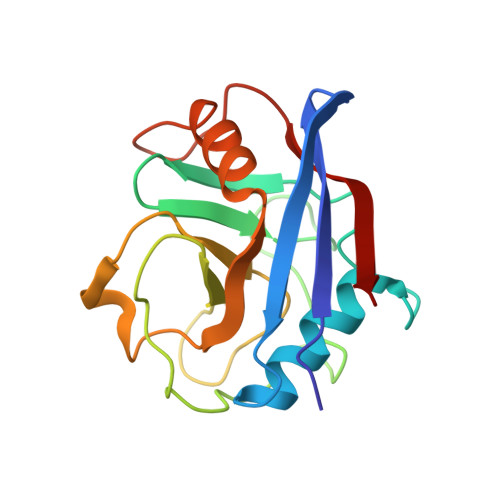
1NMK

The Sanglifehrin-Cyclophilin Interaction: Degradation Work, Synthetic Macrocyclic Analogues, X-ray Crystal Structure and Binding Data


Experimental Data Snapshot

  • Method: X-RAY DIFFRACTION
  • Resolution: 2.10 Å
  • R-Value Free: 0.189 
  • R-Value Work: 0.163 
  • R-Value Observed: 0.165 

wwPDB Validation   3D Report Full Report


Ligand Structure Quality Assessment 


This is version 1.3 of the entry. See complete history


Literature

Sanglifehrin-Cyclophilin Interaction: Degradation Work, Synthetic Macrocyclic Analogues, X-ray Crystal Structure and Binding Data

Sedrani, R.Kallen, J.Cabrejas, L.M.M.Papageorgiou, C.D.Senia, F.Rohrbach, S.Wagner, D.Thai, B.Eme, A.-M.J.France, J.Oberer, L.Rihs, G.Zenke, G.Wagner, J.

(2003) J Am Chem Soc 125: 3849-3859

  • DOI: https://doi.org/10.1021/ja021327y
  • Primary Citation of Related Structures:  
    1NMK

  • PubMed Abstract: 

    Sanglifehrin A (SFA) is a novel immunosuppressive natural product isolated from Streptomyces sp. A92-308110. SFA has a very strong affinity for cyclophilin A (IC(50) = 6.9 +/- 0.9 nM) but is structurally different from cyclosporin A (CsA) and exerts its immunosuppressive activity via a novel mechanism. SFA has a complex molecular structure consisting of a 22-membered macrocycle, bearing in position 23 a nine-carbon tether terminated by a highly substituted spirobicyclic moiety. Selective oxidative cleavage of the C(26)=C(27) exocyclic double bond affords the spirolactam containing fragment 1 and macrolide 2. The affinity of 2 for cyclophilin (IC(50) = 29 +/- 2.1 nM) is essentially identical to SFA, which indicates that the interaction between SFA and cyclophilin A is mediated exclusively by the macrocyclic portion of the molecule. This observation was confirmed by the X-ray crystal structure resolved at 2.1 A of cyclophilin A complexed to macrolide 16, a close analogue of 2. The X-ray crystal structure showed that macrolide 16 binds to the same deep hydrophobic pocket of cyclophilin A as CsA. Additional valuable details of the structure-activity relationship were obtained by two different chemical approaches: (1) degradation work on macrolide 2 or (2) synthesis of a library of macrolide analogues using the ring-closing metathesis reaction as the key step. Altogether, it appears that the complex macrocyclic fragment of SFA is a highly optimized combination of multiple functionalities including an (E,E)-diene, a short polypropionate fragment, and an unusual tripeptide unit, which together provide an extremely strong affinity for cyclophilin A.


  • Organizational Affiliation

    Transplantation Research, Novartis Pharma AG, S-507.312, CH-4002 Basel, Switzerland.


Macromolecules
Find similar proteins by:  (by identity cutoff)  |  3D Structure
Entity ID: 1
MoleculeChains Sequence LengthOrganismDetailsImage
Peptidyl-prolyl cis-trans isomerase A
A, B
165Homo sapiensMutation(s): 0 
Gene Names: PPIA OR CYPA
EC: 5.2.1.8
UniProt & NIH Common Fund Data Resources
Find proteins for P62937 (Homo sapiens)
Explore P62937 
Go to UniProtKB:  P62937
PHAROS:  P62937
GTEx:  ENSG00000196262 
Entity Groups  
Sequence Clusters30% Identity50% Identity70% Identity90% Identity95% Identity100% Identity
UniProt GroupP62937
Sequence Annotations
Expand
  • Reference Sequence
Small Molecules
Ligands 1 Unique
IDChains Name / Formula / InChI Key2D Diagram3D Interactions
SFM
Query on SFM

Download Ideal Coordinates CCD File 
C [auth A],
D [auth B]
(13E,15E)-(3S,6S,9R,10R,11S,12S,18S,21S)-10,12-DIHYDROXY-3-(3-HYDROXYBEN-ZYL)-18-((E)-3-HYDROXY-1-METHYLPROPENYL)-6-ISOPROPYL-11-METHYL-9-(3-OXO-BUTYL)-19-OXA-1,4,7,25-TETRAAZA-BICYCLO[19.3.1]PENTACOSA-13,15-DIENE-2,5,8,20-TETRAONE
C39 H56 N4 O10
WZVMDDRRINUPRQ-APBCKSDASA-N
Binding Affinity Annotations 
IDSourceBinding Affinity
SFM PDBBind:  1NMK IC50: 5.7 (nM) from 1 assay(s)
Binding MOAD:  1NMK IC50: 5.7 (nM) from 1 assay(s)
Experimental Data & Validation

Experimental Data

  • Method: X-RAY DIFFRACTION
  • Resolution: 2.10 Å
  • R-Value Free: 0.189 
  • R-Value Work: 0.163 
  • R-Value Observed: 0.165 
  • Space Group: P 21 21 21
Unit Cell:
Length ( Å )Angle ( ˚ )
a = 58.2α = 90
b = 67.3β = 90
c = 78.2γ = 90
Software Package:
Software NamePurpose
REFMACrefinement
DENZOdata reduction
SCALEPACKdata scaling
X-PLORphasing

Structure Validation

View Full Validation Report


Ligand Structure Quality Assessment 


Entry History 

Deposition Data

Revision History  (Full details and data files)

  • Version 1.0: 2003-04-01
    Type: Initial release
  • Version 1.1: 2008-04-29
    Changes: Version format compliance
  • Version 1.2: 2011-07-13
    Changes: Non-polymer description, Version format compliance
  • Version 1.3: 2023-08-16
    Changes: Data collection, Database references, Derived calculations, Refinement description