1OMP

CRYSTALLOGRAPHIC EVIDENCE OF A LARGE LIGAND-INDUCED HINGE-TWIST MOTION BETWEEN THE TWO DOMAINS OF THE MALTODEXTRIN-BINDING PROTEIN INVOLVED IN ACTIVE TRANSPORT AND CHEMOTAXIS


Experimental Data Snapshot

  • Method: X-RAY DIFFRACTION
  • Resolution: 1.80 Å
  • R-Value Work: 0.210 
  • R-Value Observed: 0.210 

wwPDB Validation   3D Report Full Report


This is version 1.5 of the entry. See complete history


Literature

Crystallographic evidence of a large ligand-induced hinge-twist motion between the two domains of the maltodextrin binding protein involved in active transport and chemotaxis.

Sharff, A.J.Rodseth, L.E.Spurlino, J.C.Quiocho, F.A.

(1992) Biochemistry 31: 10657-10663

  • DOI: https://doi.org/10.1021/bi00159a003
  • Primary Citation of Related Structures:  
    1OMP

  • PubMed Abstract: 

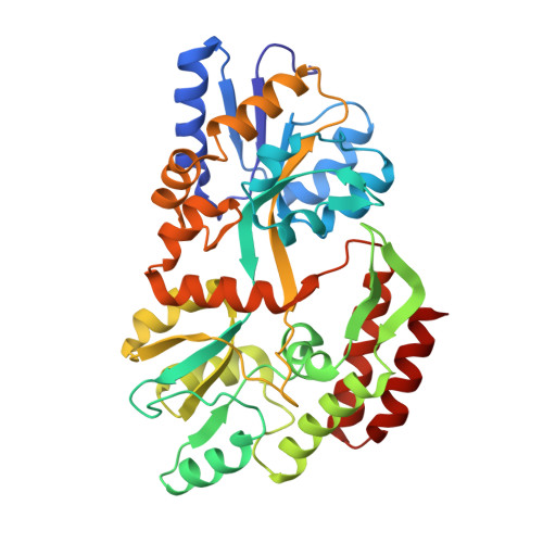
    The periplasmic maltodextrin binding protein of Escherichia coli serves as an initial receptor for the active transport of and chemotaxis toward maltooligosaccharides. The three-dimensional structure of the binding protein complexed with maltose has been previously reported [Spurlino, J. C., Lu, G.-Y., & Quiocho, F. A. (1991) J. Biol. Chem. 266, 5202-5219]. Here we report the structure of the unliganded form of the binding protein refined to 1.8-A resolution. This structure, combined with that for the liganded form, provides the first crystallographic evidence that a major ligand-induced conformational change occurs in a periplasmic binding protein. The unliganded structure shows a rigid-body "hinge-bending" between the two globular domains by approximately 35 degrees, relative to the maltose-bound structure, opening the sugar binding site groove located between the two domains. In addition, there is an 8 degrees twist of one domain relative to the other domain. The conformational changes observed between this structure and the maltose-bound structure are consistent with current models of maltose/maltodextrin transport and maltose chemotaxis and solidify a mechanism for receptor differentiation between the ligand-free and ligand-bound forms in signal transduction.


  • Organizational Affiliation

    Howard Hughes Medical Institute, Baylor College of Medicine, Houston, Texas 77030.


Macromolecules
Find similar proteins by:  (by identity cutoff)  |  3D Structure
Entity ID: 1
MoleculeChains Sequence LengthOrganismDetailsImage
D-MALTODEXTRIN BINDING PROTEIN370Escherichia coliMutation(s): 0 
UniProt
Find proteins for P0AEX9 (Escherichia coli (strain K12))
Explore P0AEX9 
Go to UniProtKB:  P0AEX9
Entity Groups  
Sequence Clusters30% Identity50% Identity70% Identity90% Identity95% Identity100% Identity
UniProt GroupP0AEX9
Sequence Annotations
Expand
  • Reference Sequence
Experimental Data & Validation

Experimental Data

  • Method: X-RAY DIFFRACTION
  • Resolution: 1.80 Å
  • R-Value Work: 0.210 
  • R-Value Observed: 0.210 
  • Space Group: P 1
Unit Cell:
Length ( Å )Angle ( ˚ )
a = 38.35α = 101
b = 44.44β = 100.4
c = 58.25γ = 104.2
Software Package:
Software NamePurpose
X-PLORmodel building
PROLSQrefinement
X-PLORrefinement
X-PLORphasing

Structure Validation

View Full Validation Report


Entry History 

Deposition Data

Revision History  (Full details and data files)

  • Version 1.0: 1994-01-31
    Type: Initial release
  • Version 1.1: 2008-03-24
    Changes: Version format compliance
  • Version 1.2: 2011-07-13
    Changes: Version format compliance
  • Version 1.3: 2019-07-17
    Changes: Data collection, Other, Refinement description
  • Version 1.4: 2019-08-14
    Changes: Data collection, Refinement description
  • Version 1.5: 2024-02-14
    Changes: Data collection, Database references