1RCA

STRUCTURE OF THE CRYSTALLINE COMPLEX OF DEOXYCYTIDYLYL-3',5'-GUANOSINE (3',5'-DCPDG) CO-CRYSTALISED WITH RIBONUCLEASE AT 1.9 ANGSTROMS RESOLUTION. RETROBINDING IN PANCREATIC RNASEA IS INDEPENDENT OF MODE OF INHIBITOR INTROMISSION


Experimental Data Snapshot

  • Method: X-RAY DIFFRACTION
  • Resolution: 1.90 Å
  • R-Value Observed: 0.218 

wwPDB Validation   3D Report Full Report


Ligand Structure Quality Assessment 


This is version 1.2 of the entry. See complete history


Literature

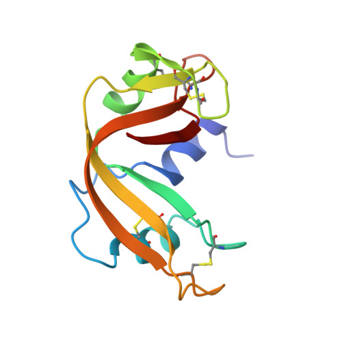
Structure of the crystalline complex of deoxycytidylyl-3',5'-guanosine (3',5'-dCpdG) cocrystallized with ribonuclease at 1.9 A resolution.

Listgarten, J.N.Maes, D.Wyns, L.Aguilar, C.F.Palmer, R.A.

(1995) Acta Crystallogr D Biol Crystallogr 51: 767-771

  • DOI: https://doi.org/10.1107/S0907444995001570
  • Primary Citation of Related Structures:  
    1RCA

  • PubMed Abstract: 

    The X-ray structure of bovine ribonuclease A cocrystallized with the dinucleotide deoxycytidylyl-3',5'-guanosine has been determined at 1.9 A resolution and refined by restrained least squares to R = 0.218 for 7807 reflections. The structure established that the recently observed retrobound mode of attachment of substrate analogues cytidylyl-2',5'-guanosine and deoxycytidylyl-3',5'-guanosine found in soaked RNase A crystals is also present in the cocrystallized complex. Retrobinding is thus unlikely to be the result of restrictions imposed by the crystalline environment as the ligands soak into the lattice but rather a phenomenon specific to small nucleotides containing guanine.


  • Organizational Affiliation

    Department of Ultrastructure, Institut voor Moleculaire Biologie, Vrije Universiteit Brussel, Sint-Genesius-Ride, Belgium.


Macromolecules
Find similar proteins by:  (by identity cutoff)  |  3D Structure
Entity ID: 1
MoleculeChains Sequence LengthOrganismDetailsImage
RIBONUCLEASE A124Bos taurusMutation(s): 0 
EC: 3.1.27.5
UniProt
Find proteins for P61823 (Bos taurus)
Explore P61823 
Go to UniProtKB:  P61823
Entity Groups  
Sequence Clusters30% Identity50% Identity70% Identity90% Identity95% Identity100% Identity
UniProt GroupP61823
Sequence Annotations
Expand
  • Reference Sequence
Experimental Data & Validation

Experimental Data

  • Method: X-RAY DIFFRACTION
  • Resolution: 1.90 Å
  • R-Value Observed: 0.218 
  • Space Group: P 1 21 1
Unit Cell:
Length ( Å )Angle ( ˚ )
a = 30.5α = 90
b = 38.7β = 106.3
c = 53.7γ = 90
Software Package:
Software NamePurpose
RESTRAINrefinement

Structure Validation

View Full Validation Report


Ligand Structure Quality Assessment 


Entry History 

Deposition Data

Revision History  (Full details and data files)

  • Version 1.0: 1995-09-15
    Type: Initial release
  • Version 1.1: 2008-03-24
    Changes: Version format compliance
  • Version 1.2: 2011-07-13
    Changes: Version format compliance