X-ray structure of a ribonuclease A-uridine vanadate complex at 1.3 A resolution.
Ladner, J.E., Wladkowski, B.D., Svensson, L.A., Sjolin, L., Gilliland, G.L.(1997) Acta Crystallogr D Biol Crystallogr 53: 290-301
- PubMed: 15299932
- DOI: https://doi.org/10.1107/S090744499601582X
- Primary Citation of Related Structures:
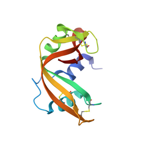
1RUV - PubMed Abstract:
The X-ray crystal structure of a uridine vanadate-ribonuclease A complex has been determined at 1.3 A resolution. The resulting structure includes all 124 amino-acid residues, a uridine vanadate, 131 water molecules, and a single bound 2-methyl-2-propanol. Side chains of 11 surface residues showing discrete disorder were modeled with multiple conformations. The final crystallographic R factor is 0.197. Structures obtained from high-level ab initio quantum calculations of model anionic oxyvanadate compounds were used to probe the effects of starting structure on the refinement process and final structure of the penta-coordinate phosphorane analog, uridine vanadate. The least-squares refinement procedure gave rise to the same final structure of the inhibitor despite significantly different starting models. Comparison with the previously determined complex of ribonuclease A with uridine vanadate obtained from a joint X-ray/neutron analysis (6RSA) [Wlodawer, Miller & Sjölin (1983). Proc. Natl Acad. Sci. USA, 80, 3628-3631] reveals similarities in the overall enzyme structure and the relative position of the key active-site residues, Hisl2, His119 and Lys41, but significant differences in the V-O bond distances and angles. The influence of ligand binding on the enzyme structure is assessed by a comparison of the current X-ray structure with the phosphate-free ribonuclease A structure (7RSA) [Wlodawer, Svensson, Sjölin & Gilliland (1988). Biochemistry, 27, 2705-2717]. Ligand binding alters the solvent structure, distribution and number of residues with multiple conformations, and temperature factors of the protein atoms. In fact, the temperature factors of atoms of several residues that interact with the ligand are reduced, but those of the atoms of several residues remote from the active site exhibit marked increases.
Organizational Affiliation:
The Center for Advanced Research in Biotechnology of the University of Maryland Biotechnology Institute and National Institute of Standards and Technology, Rockville, MD 20850, USA.