Crystal structure determination at 2.3-A resolution of human transthyretin-3',5'-dibromo-2',4,4',6-tetrahydroxyaurone complex.
Ciszak, E., Cody, V., Luft, J.R.(1992) Proc Natl Acad Sci U S A 89: 6644-6648
- PubMed: 1631168
- DOI: https://doi.org/10.1073/pnas.89.14.6644
- Primary Citation of Related Structures:
1THC - PubMed Abstract:
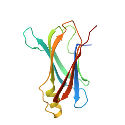
The crystal structure of the complex of 3',5'-dibromo-2',4,4',6-tetrahydroxyaurone, a flavone derivative, with human transthyretin (TTR), a serum thyroid hormone transport protein, has been determined and refined to R = 17.9% for data to 2.3-A resolution and provides a detailed description of a protein-bound flavonoid structure. This bromoaurone is a potent competitor for thyroid hormone binding to TTR, a 54,980-dalton alpha 4 tetrameric protein of 222 molecular symmetry, as well as an inhibitor of iodothyronine deiodinase. Crystals of the TTR-bromoaurone complex are isomorphous to those of native TTR. Interpretation of difference Fourier electron density maps revealed two binding modes for the bromoaurone in each of the two independent binding sites of the TTR tetramer: deep in the channel near Ser-117 (mode I) and near the channel entrance (mode II). None of the binding modes can be fully occupied because of overlap between binding positions. A statistical disorder for bromoaurone binding was also applied, as it binds along the twofold crystallographic axis and does not possess such symmetry. The binding of mode I and that of mode II were refined at half occupancy, resulting in two molecules per tetramer. The bromoaurone binds in a nonplanar antiskewed conformation. The molecular pattern for TTR binding consists of halogen groups able to anchor between beta-sheets to form both hydrophobic and hydrophilic contacts. Comparison of structural data for bromoaurone- and thyroxine-TTR complexes indicates that bromoaurone binding mode I is 3 A deeper in the channel and binding mode II is 4 A further from the channel center than thyroxine. The bromoaurone binding observed in this TTR complex differs significantly from that based upon computer modeling studies.
Organizational Affiliation:
Molecular Biophysics Department, Medical Foundation of Buffalo, Inc., NY 14203.