1U1Y

Crystal structure of a complex between WT bacteriophage MS2 coat protein and an F5 aptamer RNA stemloop with 2aminopurine substituted at the-10 position


Experimental Data Snapshot

  • Method: X-RAY DIFFRACTION
  • Resolution: 2.85 Å
  • R-Value Free: 0.196 
  • R-Value Work: 0.188 
  • R-Value Observed: 0.188 

wwPDB Validation   3D Report Full Report


This is version 1.3 of the entry. See complete history


Literature

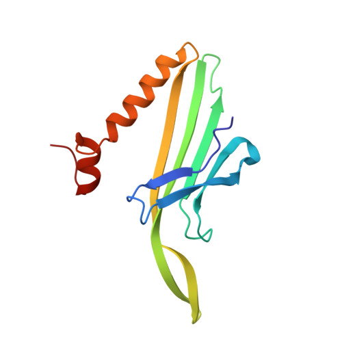
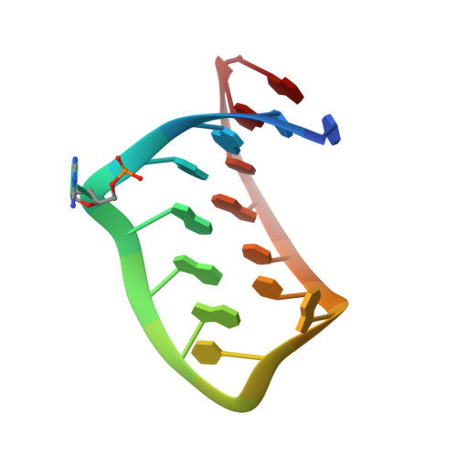
The crystal structure of a high affinity RNA stem-loop complexed with the bacteriophage MS2 capsid: further challenges in the modeling of ligand-RNA interactions.

Horn, W.T.Convery, M.A.Stonehouse, N.J.Adams, C.J.Liljas, L.Phillips, S.E.Stockley, P.G.

(2004) RNA 10: 1776-1782

  • DOI: https://doi.org/10.1261/rna.7710304
  • Primary Citation of Related Structures:  
    1U1Y

  • PubMed Abstract: 

    We have determined the structure to 2.8 A of an RNA aptamer (F5), containing 2'-deoxy-2-aminopurine (2AP) at the -10 position, complexed with MS2 coat protein by soaking the RNA into precrystallised MS2 capsids. The -10 position of the RNA is an important determinant of binding affinity for coat protein. Adenine at this position in other RNA stem-loops makes three hydrogen bonds to protein functional groups. Substituting 2AP for the -10 adenine in the F5 aptamer yields an RNA with the highest yet reported affinity for coat protein. The refined X-ray structure shows that the 2AP base makes an additional hydrogen bond to the protein compared to adenine that is presumably the principal origin of the increased affinity. There are also slight changes in phosphate backbone positions compared to unmodified F5 that probably also contribute to affinity. Such phosphate movements are common in structures of RNAs bound to the MS2 T = 3 protein shell and highlight problems for de novo design of RNA binding ligands.


  • Organizational Affiliation

    Astbury Centre for Structural Molecular Biology, University of Leeds, Leeds LS2 9JT, United Kingdom.


Macromolecules

Find similar proteins by:  (by identity cutoff)  |  3D Structure
Entity ID: 2
MoleculeChains Sequence LengthOrganismDetailsImage
Coat proteinC [auth A],
D [auth B],
E [auth C]
129Escherichia phage MS2Mutation(s): 0 
UniProt
Find proteins for P03612 (Escherichia phage MS2)
Explore P03612 
Go to UniProtKB:  P03612
Entity Groups  
Sequence Clusters30% Identity50% Identity70% Identity90% Identity95% Identity100% Identity
UniProt GroupP03612
Sequence Annotations
Expand
  • Reference Sequence

Find similar nucleic acids by:  Sequence   |   3D Structure  

Entity ID: 1
MoleculeChains LengthOrganismImage
5'-R(*CP*CP*GP*GP*(2PR)P*GP*GP*AP*UP*CP*AP*CP*CP*AP*CP*GP*G)-3'A [auth R],
B [auth S]
17N/A
Sequence Annotations
Expand
  • Reference Sequence
Experimental Data & Validation

Experimental Data

  • Method: X-RAY DIFFRACTION
  • Resolution: 2.85 Å
  • R-Value Free: 0.196 
  • R-Value Work: 0.188 
  • R-Value Observed: 0.188 
  • Space Group: H 3 2
Unit Cell:
Length ( Å )Angle ( ˚ )
a = 288.61α = 90
b = 288.61β = 90
c = 654.68γ = 120
Software Package:
Software NamePurpose
MOSFLMdata reduction
SCALAdata scaling
X-PLORmodel building
X-PLORrefinement
CCP4data scaling
X-PLORphasing

Structure Validation

View Full Validation Report


Entry History 

Deposition Data

Revision History  (Full details and data files)

  • Version 1.0: 2004-12-07
    Type: Initial release
  • Version 1.1: 2008-04-30
    Changes: Version format compliance
  • Version 1.2: 2011-07-13
    Changes: Version format compliance
  • Version 1.3: 2023-08-23
    Changes: Data collection, Database references, Derived calculations, Refinement description