Ultrafast and Low Barrier Motions in the Photoreactions of the Green Fluorescent Protein
Van Thor, J.J., Georgiev, G.Y., Towrie, M., Sage, J.T.(2005) J Biol Chem 280: 33652
- PubMed: 16033764
- DOI: https://doi.org/10.1074/jbc.M505473200
- Primary Citation of Related Structures:
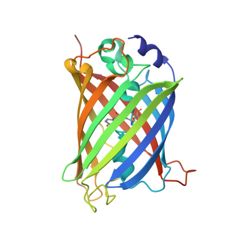
1W7S, 1W7T, 1W7U - PubMed Abstract:
Green fluorescent protein (GFP) fluoresces efficiently under blue excitation despite major electrostatic rearrangements resulting from photoionization of the chromophore and neutralization of Glu-222. A competing phototransformation process, which ionizes the chromophore and decarboxylates Glu-222, mimics the electrostatic and structural changes in the fluorescence photocycle. Structural and spectroscopic analysis of the cryogenically stabilized photoproduct at 100 K and a structurally annealed intermediate of the phototransformed protein at 170 K reveals distinct structural relaxations involving protein, chromophore, solvent, and photogenerated CO2. Strong structural changes of the 100 K photoproduct after decarboxylation appear exclusively within 15 angstroms of the chromophore and include the electrostatically driven perturbations of Gln-69, Cys-70, and water molecules in an H-bonding network connecting the chromophore. X-ray crystallography to 1.85 angstroms resolution and static and picosecond time-resolved IR spectroscopy identify structural mechanisms common to phototransformation and to the fluorescence photocycle. In particular, the appearance of a 1697 cm(-1) (+) difference band in both photocycle and phototransformation intermediates is a spectroscopic signature for the structural perturbation of Gln-69. This is taken as evidence for an electrostatically driven dynamic response that is common to both photoreaction pathways. The interactions between the chromophore and the perturbed residues and solvent are decreased or removed in the T203H single and T203H/Q69L double mutants, resulting in a strong reduction of the fluorescence quantum yield. This suggests that the electrostatic response to the transient formation of a buried charge in the wild type is important for the bright fluorescence.
Organizational Affiliation:
Laboratory of Molecular Biophysics, University of Oxford, Rex Richards Building, South Parks Road, Oxford OX1 3QU, United Kingdom. jasper@biop.ox.ac.uk