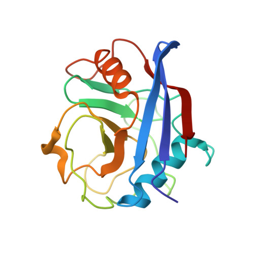
1W8M

Enzymatic and Structural Characterisation of Non Peptide Ligand Cyclophilin Complexes


Experimental Data Snapshot

  • Method: X-RAY DIFFRACTION
  • Resolution: 1.65 Å
  • R-Value Free: 0.186 
  • R-Value Work: 0.130 
  • R-Value Observed: 0.132 

wwPDB Validation   3D Report Full Report


This is version 1.3 of the entry. See complete history


Literature

Enzymatic and Structural Characterization of Non-Peptide Ligand-Cyclophilin Complexes

Kontopidis, G.Taylor, P.Walkinshaw, M.

(2004) Acta Crystallogr D Biol Crystallogr 60: 479

  • DOI: https://doi.org/10.1107/S0907444904000174
  • Primary Citation of Related Structures:  
    1W8L, 1W8M, 1W8V

  • PubMed Abstract: 

    Piperidine ligands are described that provide the first examples of non-peptidic ligand structures for the cyclophilin family of proteins. Crystal structures of two ligand complexes are compared with the unliganded protein and show ligand-induced changes in side-chain conformation and water binding. A peptidylprolyl cis-trans-isomerase assay showed the dissociation constants of the two ligands to be 320 and 25 mM. This study also provides the first published data for both enzymatic activity and three-dimensional structure for any protein-ligand complex that binds with a high-millimolar dissociation constant. The structures may be of relevance in the field of drug design, as they suggest starting points for the design of larger tighter-binding analogues.


  • Organizational Affiliation

    Structural Biochemistry Group, Department of Biochemistry, The University of Edinburgh, Michael Swann Building, King's Buildings, Edinburgh EH9 3JR, Scotland.


Macromolecules
Find similar proteins by:  (by identity cutoff)  |  3D Structure
Entity ID: 1
MoleculeChains Sequence LengthOrganismDetailsImage
PEPTIDYL-PROLYL CIS-TRANS ISOMERASE A165Homo sapiensMutation(s): 0 
EC: 5.2.1.8
UniProt & NIH Common Fund Data Resources
Find proteins for P62937 (Homo sapiens)
Explore P62937 
Go to UniProtKB:  P62937
PHAROS:  P62937
GTEx:  ENSG00000196262 
Entity Groups  
Sequence Clusters30% Identity50% Identity70% Identity90% Identity95% Identity100% Identity
UniProt GroupP62937
Sequence Annotations
Expand
  • Reference Sequence
Small Molecules
Ligands 1 Unique
IDChains Name / Formula / InChI Key2D Diagram3D Interactions
E1P
Query on E1P

Download Ideal Coordinates CCD File 
B [auth A]ETHYL OXO(PIPERIDIN-1-YL)ACETATE
C9 H15 N O3
ASBDXHCMVYVJQQ-UHFFFAOYSA-N
Binding Affinity Annotations 
IDSourceBinding Affinity
E1P Binding MOAD:  1W8M Ki: 2.50e+7 (nM) from 1 assay(s)
PDBBind:  1W8M Ki: 2.50e+7 (nM) from 1 assay(s)
Experimental Data & Validation

Experimental Data

  • Method: X-RAY DIFFRACTION
  • Resolution: 1.65 Å
  • R-Value Free: 0.186 
  • R-Value Work: 0.130 
  • R-Value Observed: 0.132 
  • Space Group: P 21 21 21
Unit Cell:
Length ( Å )Angle ( ˚ )
a = 36.307α = 90
b = 54.536β = 90
c = 71.099γ = 90
Software Package:
Software NamePurpose
REFMACrefinement
DENZOdata reduction
SCALEPACKdata scaling
X-PLORphasing

Structure Validation

View Full Validation Report


Entry History 

Deposition Data

Revision History  (Full details and data files)

  • Version 1.0: 2004-09-30
    Type: Initial release
  • Version 1.1: 2011-05-08
    Changes: Version format compliance
  • Version 1.2: 2011-07-13
    Changes: Version format compliance
  • Version 1.3: 2023-12-13
    Changes: Data collection, Database references, Refinement description