2AX2

Production and X-ray crystallographic analysis of fully deuterated human carbonic anhydrase II


Experimental Data Snapshot

  • Method: X-RAY DIFFRACTION
  • Resolution: 1.50 Å
  • R-Value Free: 0.206 
  • R-Value Work: 0.195 

wwPDB Validation   3D Report Full Report


This is version 1.3 of the entry. See complete history


Literature

Production and X-ray crystallographic analysis of fully deuterated human carbonic anhydrase II.

Budayova-Spano, M.Fisher, S.Z.Dauvergne, M.T.Agbandje-McKenna, M.Silverman, D.N.Myles, D.A.McKenna, R.

(2006) Acta Crystallogr Sect F Struct Biol Cryst Commun 62: 6-9

  • DOI: https://doi.org/10.1107/S1744309105038248
  • Primary Citation of Related Structures:  
    2AX2

  • PubMed Abstract: 

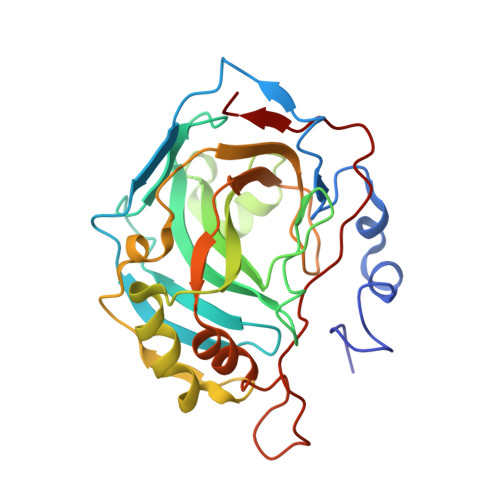
    Human carbonic anhydrase II (HCA II) is a zinc metalloenzyme that catalyzes the reversible hydration and dehydration of carbon dioxide and bicarbonate, respectively. The rate-limiting step in catalysis is the intramolecular transfer of a proton between the zinc-bound solvent (H2O/OH-) and the proton-shuttling residue His64. This distance (approximately 7.5 A) is spanned by a well defined active-site solvent network stabilized by amino-acid side chains (Tyr7, Asn62, Asn67, Thr199 and Thr200). Despite the availability of high-resolution (approximately 1.0 A) X-ray crystal structures of HCA II, there is currently no definitive information available on the positions and orientations of the H atoms of the solvent network or active-site amino acids and their ionization states. In preparation for neutron diffraction studies to elucidate this hydrogen-bonding network, perdeuterated HCA II has been expressed, purified, crystallized and its X-ray structure determined to 1.5 A resolution. The refined structure is highly isomorphous with hydrogenated HCA II, especially with regard to the active-site architecture and solvent network. This work demonstrates the suitability of these crystals for neutron macromolecular crystallography.


  • Organizational Affiliation

    European Molecular Biology Laboratory Grenoble Outstation, 6 Rue Jules Horowitz, 38042 Grenoble, France.


Macromolecules
Find similar proteins by:  (by identity cutoff)  |  3D Structure
Entity ID: 1
MoleculeChains Sequence LengthOrganismDetailsImage
Carbonic anhydrase II260Homo sapiensMutation(s): 0 
Gene Names: CA2
EC: 4.2.1.1
UniProt & NIH Common Fund Data Resources
Find proteins for P00918 (Homo sapiens)
Explore P00918 
Go to UniProtKB:  P00918
PHAROS:  P00918
GTEx:  ENSG00000104267 
Entity Groups  
Sequence Clusters30% Identity50% Identity70% Identity90% Identity95% Identity100% Identity
UniProt GroupP00918
Sequence Annotations
Expand
  • Reference Sequence
Small Molecules
Ligands 1 Unique
IDChains Name / Formula / InChI Key2D Diagram3D Interactions
ZN
Query on ZN

Download Ideal Coordinates CCD File 
B [auth A]ZINC ION
Zn
PTFCDOFLOPIGGS-UHFFFAOYSA-N
Experimental Data & Validation

Experimental Data

  • Method: X-RAY DIFFRACTION
  • Resolution: 1.50 Å
  • R-Value Free: 0.206 
  • R-Value Work: 0.195 
  • Space Group: P 1 21 1
Unit Cell:
Length ( Å )Angle ( ˚ )
a = 42.149α = 90
b = 41.124β = 104.37
c = 72.014γ = 90
Software Package:
Software NamePurpose
DENZOdata reduction
SCALEPACKdata scaling
CNSrefinement
CNSphasing

Structure Validation

View Full Validation Report


Entry History 

Deposition Data

Revision History  (Full details and data files)

  • Version 1.0: 2006-01-03
    Type: Initial release
  • Version 1.1: 2008-05-01
    Changes: Version format compliance
  • Version 1.2: 2011-07-13
    Changes: Version format compliance
  • Version 1.3: 2023-08-23
    Changes: Data collection, Database references, Derived calculations, Refinement description