2BCX

Crystal structure of calmodulin in complex with a ryanodine receptor peptide


Experimental Data Snapshot

  • Method: X-RAY DIFFRACTION
  • Resolution: 2.00 Å
  • R-Value Free: 0.249 
  • R-Value Work: 0.213 
  • R-Value Observed: 0.214 

wwPDB Validation   3D Report Full Report


This is version 1.4 of the entry. See complete history


Literature

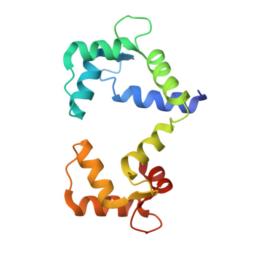
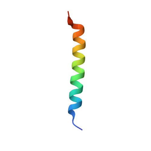
Complex of calmodulin with a ryanodine receptor target reveals a novel, flexible binding mode.

Maximciuc, A.A.Putkey, J.A.Shamoo, Y.Mackenzie, K.R.

(2006) Structure 14: 1547-1556

  • DOI: https://doi.org/10.1016/j.str.2006.08.011
  • Primary Citation of Related Structures:  
    2BCX

  • PubMed Abstract: 

    Calmodulin regulates ryanodine receptor-mediated Ca(2+) release through a conserved binding site. The crystal structure of Ca(2+)-calmodulin bound to this conserved site reveals that calmodulin recognizes two hydrophobic anchor residues at a novel "1-17" spacing that brings the calmodulin lobes close together but prevents them from contacting one another. NMR residual dipolar couplings demonstrate that the detailed structure of each lobe is preserved in solution but also show that the lobes experience domain motions within the complex. FRET measurements confirm the close approach of the lobes in binding the 1-17 target and show that calmodulin binds with one lobe to a peptide lacking the second anchor. We suggest that calmodulin regulates the Ca(2+) channel by switching between the contiguous binding mode seen in our crystal structure and a state where one lobe of calmodulin contacts the conserved binding site while the other interacts with a noncontiguous site on the channel.


  • Organizational Affiliation

    Department of Biochemistry and Cell Biology, Rice University, Houston, Texas 77005, USA.


Macromolecules
Find similar proteins by:  (by identity cutoff)  |  3D Structure
Entity ID: 1
MoleculeChains Sequence LengthOrganismDetailsImage
Calmodulin148Gallus gallusMutation(s): 0 
Gene Names: CALM
UniProt
Find proteins for P62149 (Gallus gallus)
Explore P62149 
Go to UniProtKB:  P62149
Entity Groups  
Sequence Clusters30% Identity50% Identity70% Identity90% Identity95% Identity100% Identity
UniProt GroupP62149
Sequence Annotations
Expand
  • Reference Sequence
Find similar proteins by:  (by identity cutoff)  |  3D Structure
Entity ID: 2
MoleculeChains Sequence LengthOrganismDetailsImage
Ryanodine receptor 130N/AMutation(s): 0 
UniProt
Find proteins for P11716 (Oryctolagus cuniculus)
Explore P11716 
Go to UniProtKB:  P11716
Entity Groups  
Sequence Clusters30% Identity50% Identity70% Identity90% Identity95% Identity100% Identity
UniProt GroupP11716
Sequence Annotations
Expand
  • Reference Sequence
Experimental Data & Validation

Experimental Data

  • Method: X-RAY DIFFRACTION
  • Resolution: 2.00 Å
  • R-Value Free: 0.249 
  • R-Value Work: 0.213 
  • R-Value Observed: 0.214 
  • Space Group: P 21 21 21
Unit Cell:
Length ( Å )Angle ( ˚ )
a = 38.1α = 90
b = 44.1β = 90
c = 90.3γ = 90
Software Package:
Software NamePurpose
d*TREKdata scaling
SHARPphasing
SOLOMONphasing
CNSrefinement
PDB_EXTRACTdata extraction

Structure Validation

View Full Validation Report


Entry History 

Deposition Data

Revision History  (Full details and data files)

  • Version 1.0: 2006-10-31
    Type: Initial release
  • Version 1.1: 2008-05-01
    Changes: Version format compliance
  • Version 1.2: 2011-07-13
    Changes: Version format compliance
  • Version 1.3: 2017-10-18
    Changes: Refinement description
  • Version 1.4: 2024-02-14
    Changes: Data collection, Database references, Derived calculations