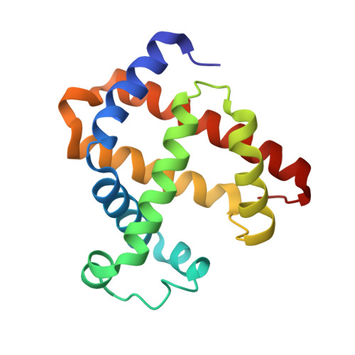
Ligand migration pathway and protein dynamics in myoglobin: a time-resolved crystallographic study on L29W MbCO.
Schmidt, M., Nienhaus, K., Pahl, R., Krasselt, A., Anderson, S., Parak, F., Nienhaus, G.U., Srajer, V.(2005) Proc Natl Acad Sci U S A 102: 11704-11709
- PubMed: 16085709
- DOI: https://doi.org/10.1073/pnas.0504932102
- Primary Citation of Related Structures:
2BW9, 2BWH - PubMed Abstract:
By using time-resolved x-ray crystallography at room temperature, structural relaxations and ligand migration were examined in myoglobin (Mb) mutant L29W from nanoseconds to seconds after photodissociation of carbon monoxide (CO) from the heme iron by nanosecond laser pulses. The data were analyzed in terms of transient kinetics by fitting trial functions to integrated difference electron density values obtained from select structural moieties, thus allowing a quantitative description of the processes involved. The observed relaxations are linked to other investigations on protein dynamics. At the earliest times, the heme has already completely relaxed into its domed deoxy structure, and there is no photo-dissociated CO visible at the primary docking site. Initial relaxations of larger globin moieties are completed within several hundred nanoseconds. They influence the concomitant migration of photo-dissociated CO to the Xe1 site, where it appears at approximately 300 ns and leaves again at approximately 1.5 ms. The extremely long residence time in Xe1 as compared with wild-type MbCO implies that, in the latter protein, the CO exits the protein from Xe1 predominantly via the distal pocket. A well-defined deligated state is populated between approximately 2 micros and approximately 1 ms; its structure is very similar to the equilibrium deoxy structure. Between 1.5 and 20 ms, no CO is visible in the protein interior; it is either distributed among many sites within the protein or has escaped to the solvent. Finally, recombination at the heme iron occurs after >20 ms.
Organizational Affiliation:
Physikdepartment E17, Technische Universität München, James Franck Strasse, 85747 Garching, Germany. marius.schmidt@ph.tum.de