2ERZ

Crystal Structure of c-AMP Dependent Kinase (PKA) bound to hydroxyfasudil


Experimental Data Snapshot

  • Method: X-RAY DIFFRACTION
  • Resolution: 2.20 Å
  • R-Value Free: 0.287 
  • R-Value Work: 0.223 
  • R-Value Observed: 0.227 

wwPDB Validation   3D Report Full Report


Ligand Structure Quality Assessment 


This is version 1.3 of the entry. See complete history


Literature

The Structure of Dimeric ROCK I Reveals the Mechanism for Ligand Selectivity.

Jacobs, M.Hayakawa, K.Swenson, L.Bellon, S.Fleming, M.Taslimi, P.Doran, J.

(2006) J Biol Chem 281: 260-268

  • DOI: https://doi.org/10.1074/jbc.M508847200
  • Primary Citation of Related Structures:  
    2ERZ, 2ESM, 2ETK, 2ETR, 3D9V

  • PubMed Abstract: 

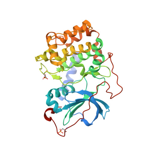
    ROCK or Rho-associated kinase, a serine/threonine kinase, is an effector of Rho-dependent signaling and is involved in actin-cytoskeleton assembly and cell motility and contraction. The ROCK protein consists of several domains: an N-terminal region, a kinase catalytic domain, a coiled-coil domain containing a RhoA binding site, and a pleckstrin homology domain. The C-terminal region of ROCK binds to and inhibits the kinase catalytic domains, and this inhibition is reversed by binding RhoA, a small GTPase. Here we present the structure of the N-terminal region and the kinase domain. In our structure, two N-terminal regions interact to form a dimerization domain linking two kinase domains together. This spatial arrangement presents the kinase active sites and regulatory sequences on a common face affording the possibility of both kinases simultaneously interacting with a dimeric inhibitory domain or with a dimeric substrate. The kinase domain adopts a catalytically competent conformation; however, no phosphorylation of active site residues is observed in the structure. We also determined the structures of ROCK bound to four different ATP-competitive small molecule inhibitors (Y-27632, fasudil, hydroxyfasudil, and H-1152P). Each of these compounds binds with reduced affinity to cAMP-dependent kinase (PKA), a highly homologous kinase. Subtle differences exist between the ROCK- and PKA-bound conformations of the inhibitors that suggest that interactions with a single amino acid of the active site (Ala215 in ROCK and Thr183 in PKA) determine the relative selectivity of these compounds. Hydroxyfasudil, a metabolite of fasudil, may be selective for ROCK over PKA through a reversed binding orientation.


  • Organizational Affiliation

    Vertex Pharmaceuticals Incorporated, Cambridge, Massachusetts 02139, USA. marc_jacobs@vrtx.com


Macromolecules
Find similar proteins by:  (by identity cutoff)  |  3D Structure
Entity ID: 1
MoleculeChains Sequence LengthOrganismDetailsImage
cAMP-dependent protein kinase, alpha-catalytic subunitA [auth E]351Mus musculusMutation(s): 2 
Gene Names: PrkacaPkaca
EC: 2.7.1.37
UniProt
Find proteins for P05132 (Mus musculus)
Explore P05132 
Go to UniProtKB:  P05132
Entity Groups  
Sequence Clusters30% Identity50% Identity70% Identity90% Identity95% Identity100% Identity
UniProt GroupP05132
Sequence Annotations
Expand
  • Reference Sequence

Find similar proteins by:  Sequence   |   3D Structure  

Entity ID: 2
MoleculeChains Sequence LengthOrganismDetailsImage
cAMP-dependent protein kinase inhibitor, alpha formB [auth I]20N/AMutation(s): 0 
UniProt
Find proteins for P61926 (Oryctolagus cuniculus)
Explore P61926 
Go to UniProtKB:  P61926
Entity Groups  
Sequence Clusters30% Identity50% Identity70% Identity90% Identity95% Identity100% Identity
UniProt GroupP61926
Sequence Annotations
Expand
  • Reference Sequence
Small Molecules
Ligands 1 Unique
IDChains Name / Formula / InChI Key2D Diagram3D Interactions
HFS
Query on HFS

Download Ideal Coordinates CCD File 
C [auth E]1-(1-HYDROXY-5-ISOQUINOLINESULFONYL)HOMOPIPERAZINE
C14 H17 N3 O3 S
ZAVGJDAFCZAWSZ-UHFFFAOYSA-N
Modified Residues  2 Unique
IDChains TypeFormula2D DiagramParent
SEP
Query on SEP
A [auth E]L-PEPTIDE LINKINGC3 H8 N O6 PSER
TPO
Query on TPO
A [auth E]L-PEPTIDE LINKINGC4 H10 N O6 PTHR
Binding Affinity Annotations 
IDSourceBinding Affinity
HFS Binding MOAD:  2ERZ Ki: 2200 (nM) from 1 assay(s)
PDBBind:  2ERZ Ki: 2200 (nM) from 1 assay(s)
Experimental Data & Validation

Experimental Data

  • Method: X-RAY DIFFRACTION
  • Resolution: 2.20 Å
  • R-Value Free: 0.287 
  • R-Value Work: 0.223 
  • R-Value Observed: 0.227 
  • Space Group: P 21 21 21
Unit Cell:
Length ( Å )Angle ( ˚ )
a = 72.794α = 90
b = 75.751β = 90
c = 80.048γ = 90
Software Package:
Software NamePurpose
d*TREKdata processing
CNSrefinement
PDB_EXTRACTdata extraction
CrystalCleardata reduction
d*TREKdata scaling
AMoREphasing
CNXrefinement

Structure Validation

View Full Validation Report


Ligand Structure Quality Assessment 


Entry History 

Deposition Data

  • Released Date: 2005-11-08 
  • Deposition Author(s): Jacobs, M.

Revision History  (Full details and data files)

  • Version 1.0: 2005-11-08
    Type: Initial release
  • Version 1.1: 2008-05-01
    Changes: Version format compliance
  • Version 1.2: 2011-07-13
    Changes: Version format compliance
  • Version 1.3: 2023-08-23
    Changes: Data collection, Database references, Derived calculations, Refinement description