2FOT

Crystal structure of the complex between calmodulin and alphaII-spectrin


Experimental Data Snapshot

  • Method: X-RAY DIFFRACTION
  • Resolution: 2.45 Å
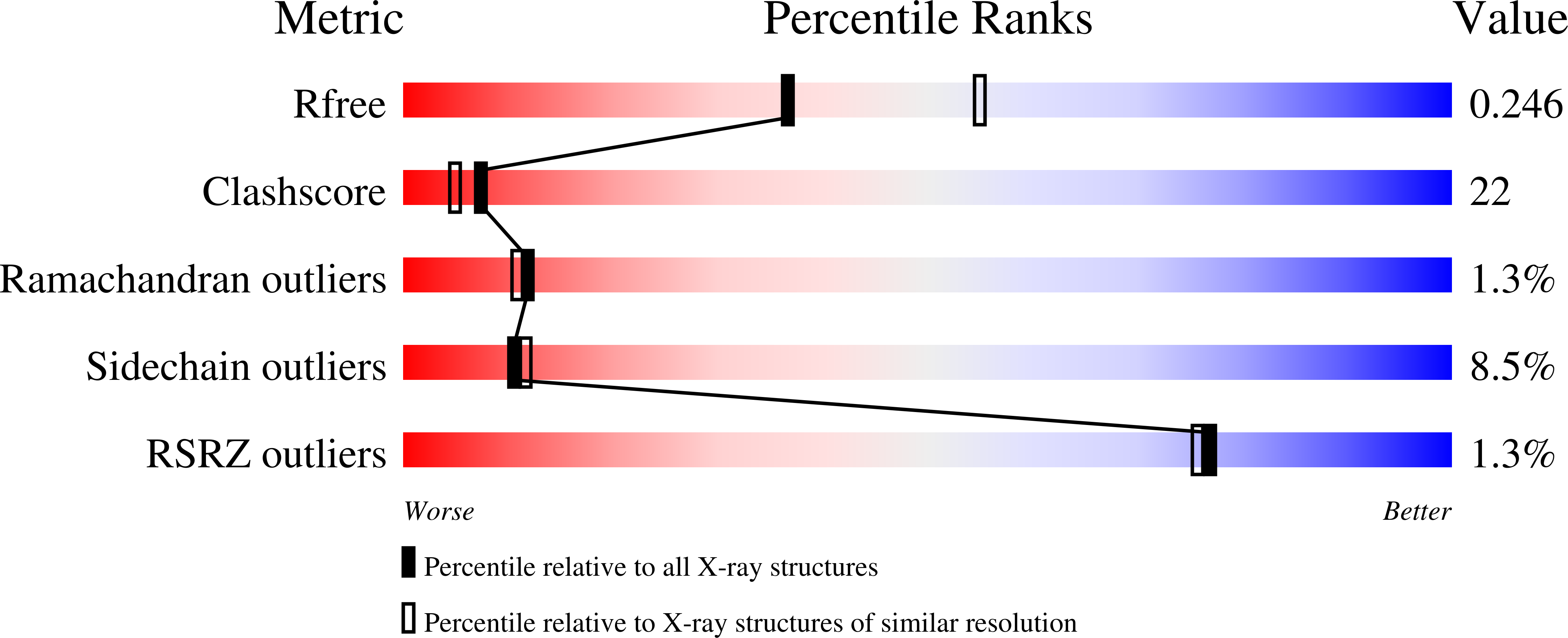
  • R-Value Free: 0.269 
  • R-Value Work: 0.245 
  • R-Value Observed: 0.245 

wwPDB Validation   3D Report Full Report


This is version 1.4 of the entry. See complete history


Literature

Structure of the calmodulin alphaII-spectrin complex provides insight into the regulation of cell plasticity.

Simonovic, M.Zhang, Z.Cianci, C.D.Steitz, T.A.Morrow, J.S.

(2006) J Biol Chem 281: 34333-34340

  • DOI: https://doi.org/10.1074/jbc.M604613200
  • Primary Citation of Related Structures:  
    2FOT

  • PubMed Abstract: 

    AlphaII-spectrin is a major cortical cytoskeletal protein contributing to membrane organization and integrity. The Ca2+-activated binding of calmodulin to an unstructured insert in the 11th repeat unit of alphaII-spectrin enhances the susceptibility of spectrin to calpain cleavage but abolishes its sensitivity to several caspases and to at least one bacterially derived pathologic protease. Other regulatory inputs including phosphorylation by c-Src also modulate the proteolytic susceptibility of alphaII-spectrin. These pathways, acting through spectrin, appear to control membrane plasticity and integrity in several cell types. To provide a structural basis for understanding these crucial biological events, we have solved the crystal structure of a complex between bovine calmodulin and the calmodulin-binding domain of human alphaII-spectrin (Protein Data Bank ID code 2FOT). The structure revealed that the entire calmodulin-spectrin-binding interface is hydrophobic in nature. The spectrin domain is also unique in folding into an amphiphilic helix once positioned within the calmodulin-binding groove. The structure of this complex provides insight into the mechanisms by which calmodulin, calpain, caspase, and tyrosine phosphorylation act on spectrin to regulate essential cellular processes.


  • Organizational Affiliation

    Howard Hughes Medical Institute, Yale University, New Haven, Connecticut 06520, USA.


Macromolecules
Find similar proteins by:  (by identity cutoff)  |  3D Structure
Entity ID: 1
MoleculeChains Sequence LengthOrganismDetailsImage
Calmodulin148Bos taurusMutation(s): 0 
UniProt
Find proteins for P62157 (Bos taurus)
Explore P62157 
Go to UniProtKB:  P62157
Entity Groups  
Sequence Clusters30% Identity50% Identity70% Identity90% Identity95% Identity100% Identity
UniProt GroupP62157
Sequence Annotations
Expand
  • Reference Sequence
Find similar proteins by:  (by identity cutoff)  |  3D Structure
Entity ID: 2
MoleculeChains Sequence LengthOrganismDetailsImage
alpha-II spectrin SpectrinB [auth C]42Homo sapiensMutation(s): 1 
Gene Names: SPTAN1SPTA2
UniProt & NIH Common Fund Data Resources
Find proteins for Q13813 (Homo sapiens)
Explore Q13813 
Go to UniProtKB:  Q13813
PHAROS:  Q13813
GTEx:  ENSG00000197694 
Entity Groups  
Sequence Clusters30% Identity50% Identity70% Identity90% Identity95% Identity100% Identity
UniProt GroupQ13813
Sequence Annotations
Expand
  • Reference Sequence
Experimental Data & Validation

Experimental Data

  • Method: X-RAY DIFFRACTION
  • Resolution: 2.45 Å
  • R-Value Free: 0.269 
  • R-Value Work: 0.245 
  • R-Value Observed: 0.245 
  • Space Group: P 21 21 21
Unit Cell:
Length ( Å )Angle ( ˚ )
a = 44.288α = 90
b = 57.737β = 90
c = 69.753γ = 90
Software Package:
Software NamePurpose
CNSrefinement
DENZOdata reduction
SCALEPACKdata scaling
PHASERphasing

Structure Validation

View Full Validation Report


Entry History 

Deposition Data

Revision History  (Full details and data files)

  • Version 1.0: 2006-09-05
    Type: Initial release
  • Version 1.1: 2008-05-01
    Changes: Version format compliance
  • Version 1.2: 2011-07-13
    Changes: Version format compliance
  • Version 1.3: 2021-10-20
    Changes: Database references, Derived calculations
  • Version 1.4: 2023-08-30
    Changes: Data collection, Refinement description