2YKS

PENTAMERIC LIGAND GATED ION CHANNEL ELIC MUTANT F246A


Experimental Data Snapshot

  • Method: X-RAY DIFFRACTION
  • Resolution: 3.30 Å
  • R-Value Free: 0.265 
  • R-Value Work: 0.242 
  • R-Value Observed: 0.243 

wwPDB Validation   3D Report Full Report


This is version 1.2 of the entry. See complete history


Literature

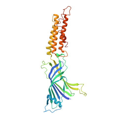
Ligand Activation of the Prokaryotic Pentameric Ligand-Gated Ion Channel Elic.

Zimmermann, I.Dutzler, R.

(2011) PLoS Biol 9: 1101

  • DOI: https://doi.org/10.1371/journal.pbio.1001101
  • Primary Citation of Related Structures:  
    2YKS

  • PubMed Abstract: 

    While the pentameric ligand-gated ion channel ELIC has recently provided first insight into the architecture of the family at high resolution, its detailed investigation was so far prevented by the fact that activating ligands were unknown. Here we describe a study on the functional characterization of ELIC by electrophysiology and X-ray crystallography. ELIC is activated by a class of primary amines that include the neurotransmitter GABA at high micro- to millimolar concentrations. The ligands bind to a conserved site and evoke currents that slowly desensitize over time. The protein forms cation selective channels with properties that resemble the nicotinic acetylcholine receptor. The high single channel conductance and the comparably simple functional behavior make ELIC an attractive model system to study general mechanisms of ion conduction and gating in this important family of neurotransmitter receptors.


  • Organizational Affiliation

    Department of Biochemistry, University of Zurich, Zurich, Switzerland.


Macromolecules
Find similar proteins by:  (by identity cutoff)  |  3D Structure
Entity ID: 1
MoleculeChains Sequence LengthOrganismDetailsImage
CYS-LOOP LIGAND-GATED ION CHANNEL
A, B, C, D, E
A, B, C, D, E, F, G, H, I, J
321Dickeya chrysanthemiMutation(s): 1 
Membrane Entity: Yes 
UniProt
Find proteins for P0C7B7 (Dickeya chrysanthemi)
Explore P0C7B7 
Go to UniProtKB:  P0C7B7
Entity Groups  
Sequence Clusters30% Identity50% Identity70% Identity90% Identity95% Identity100% Identity
UniProt GroupP0C7B7
Sequence Annotations
Expand
  • Reference Sequence
Experimental Data & Validation

Experimental Data

  • Method: X-RAY DIFFRACTION
  • Resolution: 3.30 Å
  • R-Value Free: 0.265 
  • R-Value Work: 0.242 
  • R-Value Observed: 0.243 
  • Space Group: P 1 21 1
Unit Cell:
Length ( Å )Angle ( ˚ )
a = 105.502α = 90
b = 266.147β = 109.52
c = 110.851γ = 90
Software Package:
Software NamePurpose
PHENIXrefinement
XDSdata reduction
XDSdata scaling
PHENIXphasing

Structure Validation

View Full Validation Report


Entry History 

Deposition Data

Revision History  (Full details and data files)

  • Version 1.0: 2011-07-06
    Type: Initial release
  • Version 1.1: 2011-11-16
    Changes: Database references, Version format compliance
  • Version 1.2: 2023-12-20
    Changes: Data collection, Database references, Other, Refinement description