2YN6

Pentameric Ligand-Gated Ion Channel ELIC in Complex with Barium


Experimental Data Snapshot

  • Method: X-RAY DIFFRACTION
  • Resolution: 3.31 Å
  • R-Value Free: 0.258 
  • R-Value Work: 0.227 
  • R-Value Observed: 0.229 

wwPDB Validation   3D Report Full Report


This is version 1.1 of the entry. See complete history


Literature

Inhibition of the Prokaryotic Pentameric Ligand-Gated Ion Channel Elic by Divalent Cations.

Zimmermann, I.Marabelli, A.Bertozzi, C.Sivilotti, L.G.Dutzler, R.

(2012) PLoS Biol 10: 1429

  • DOI: https://doi.org/10.1371/journal.pbio.1001429
  • Primary Citation of Related Structures:  
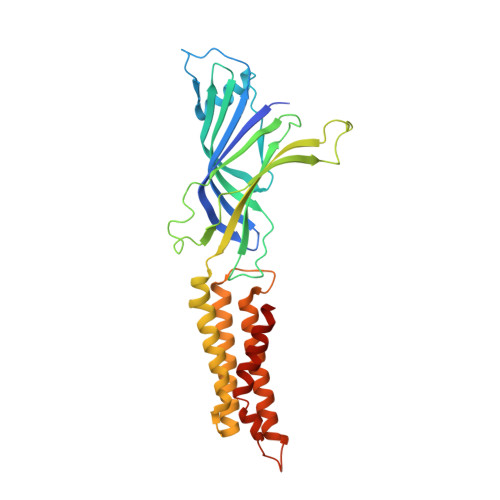
    2YN6

  • PubMed Abstract: 

    The modulation of pentameric ligand-gated ion channels (pLGICs) by divalent cations is believed to play an important role in their regulation in a physiological context. Ions such as calcium or zinc influence the activity of pLGIC neurotransmitter receptors by binding to their extracellular domain and either potentiate or inhibit channel activation. Here we have investigated by electrophysiology and X-ray crystallography the effect of divalent ions on ELIC, a close prokaryotic pLGIC homologue of known structure. We found that divalent cations inhibit the activation of ELIC by the agonist cysteamine, reducing both its potency and, at higher concentrations, its maximum response. Crystal structures of the channel in complex with barium reveal the presence of several distinct binding sites. By mutagenesis we confirmed that the site responsible for divalent inhibition is located at the outer rim of the extracellular domain, at the interface between adjacent subunits but at some distance from the agonist binding region. Here, divalent cations interact with the protein via carboxylate side-chains, and the site is similar in structure to calcium binding sites described in other proteins. There is evidence that other pLGICs may be regulated by divalent ions binding to a similar region, even though the interacting residues are not conserved within the family. Our study provides structural and functional insight into the allosteric regulation of ELIC and is of potential relevance for the entire family.


  • Organizational Affiliation

    Department of Biochemistry, University of Zürich, Zürich, Switzerland.


Macromolecules
Find similar proteins by:  (by identity cutoff)  |  3D Structure
Entity ID: 1
MoleculeChains Sequence LengthOrganismDetailsImage
PENTAMERIC LIGAND-GATED ION CHANNEL ELIC
A, B, C, D, E
322Dickeya dadantii 3937Mutation(s): 0 
Membrane Entity: Yes 
UniProt
Find proteins for E0SJQ4 (Dickeya dadantii (strain 3937))
Explore E0SJQ4 
Go to UniProtKB:  E0SJQ4
Entity Groups  
Sequence Clusters30% Identity50% Identity70% Identity90% Identity95% Identity100% Identity
UniProt GroupE0SJQ4
Sequence Annotations
Expand
  • Reference Sequence
Small Molecules
Ligands 1 Unique
IDChains Name / Formula / InChI Key2D Diagram3D Interactions
BA
Query on BA

Download Ideal Coordinates CCD File 
F [auth A]
G [auth A]
H [auth B]
I [auth B]
J [auth C]
F [auth A],
G [auth A],
H [auth B],
I [auth B],
J [auth C],
K [auth C],
L [auth C],
M [auth D],
N [auth E],
O [auth E],
P [auth E],
Q [auth E]
BARIUM ION
Ba
XDFCIPNJCBUZJN-UHFFFAOYSA-N
Experimental Data & Validation

Experimental Data

  • Method: X-RAY DIFFRACTION
  • Resolution: 3.31 Å
  • R-Value Free: 0.258 
  • R-Value Work: 0.227 
  • R-Value Observed: 0.229 
  • Space Group: P 43
Unit Cell:
Length ( Å )Angle ( ˚ )
a = 100.473α = 90
b = 100.473β = 90
c = 263.951γ = 90
Software Package:
Software NamePurpose
PHENIXrefinement
XDSdata reduction
XSCALEdata scaling
PHASERphasing

Structure Validation

View Full Validation Report


Entry History 

Deposition Data

Revision History  (Full details and data files)

  • Version 1.0: 2012-12-12
    Type: Initial release
  • Version 1.1: 2023-12-20
    Changes: Data collection, Database references, Derived calculations, Other, Refinement description