


2ZFE
Crystal structure of bacteriorhodopsin-xenon complex
- PDB DOI: https://doi.org/10.2210/pdb2ZFE/pdb
- Classification: PROTON TRANSPORT
- Organism(s): Halobacterium salinarum (strain ATCC 700922 / JCM 11081 / NRC-1)
- Mutation(s): No
- Membrane Protein: Yes PDBTMMemProtMD
- Deposited: 2007-12-31 Released: 2008-11-11
Experimental Data Snapshot
- Method: X-RAY DIFFRACTION
- Resolution: 2.50 Å
- R-Value Free: 0.279
- R-Value Work: 0.250
- R-Value Observed: 0.250

This is version 2.1 of the entry. See complete history.

Macromolecules
Find similar proteins by:
(by identity cutoff) | 3D Structure
Entity ID: 1 | |||||
|---|---|---|---|---|---|
| Molecule | Chains | Sequence Length | Organism | Details | Image |
| Bacteriorhodopsin | 262 | N/A | Mutation(s): 0 Membrane Entity: Yes | 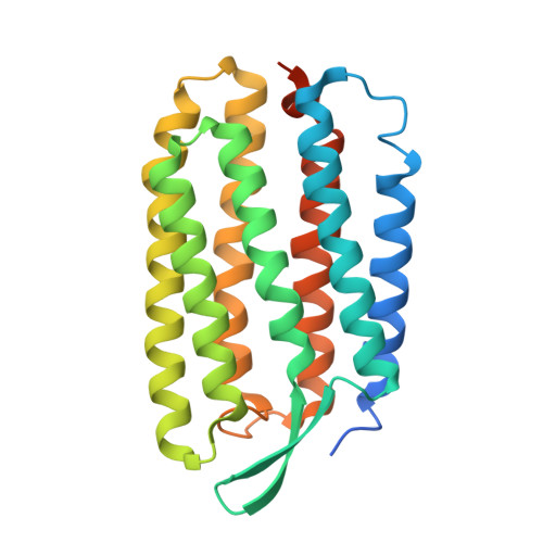 | |
UniProt | |||||
Find proteins for P02945 (Halobacterium salinarum (strain ATCC 700922 / JCM 11081 / NRC-1)) Explore P02945 Go to UniProtKB: P02945 | |||||
Entity Groups | |||||
| Sequence Clusters | 30% Identity50% Identity70% Identity90% Identity95% Identity100% Identity | ||||
| UniProt Group | P02945 | ||||
Sequence AnnotationsExpand | |||||
| |||||
Oligosaccharides

Small Molecules
| Ligands 5 Unique | |||||
|---|---|---|---|---|---|
| ID | Chains | Name / Formula / InChI Key | 2D Diagram | 3D Interactions | |
| L3P Query on L3P | D [auth A] | 2,3-DI-O-PHYTANLY-3-SN-GLYCERO-1-PHOSPHORYL-3'-SN-GLYCEROL-1'-PHOSPHATE C46 H94 O11 P2 TZXJQSKPTCRGCA-VZSPAKCESA-L |  | ||
| L1P Query on L1P | F [auth A], G [auth A], H [auth A] | 3-PHOSPHORYL-[1,2-DI-PHYTANYL]GLYCEROL C43 H89 O6 P UKQGAMWGTOTQPC-ALOLAALWSA-N |  | ||
| L2P Query on L2P | E [auth A] | 2,3-DI-PHYTANYL-GLYCEROL C43 H88 O3 ISDBCJSGCHUHFI-UMZPFTBHSA-N |  | ||
| RET Query on RET | C [auth A] | RETINAL C20 H28 O NCYCYZXNIZJOKI-OVSJKPMPSA-N |  | ||
| XE Query on XE | I [auth A] | XENON Xe FHNFHKCVQCLJFQ-UHFFFAOYSA-N |  | ||
Experimental Data & Validation
Experimental Data
- Method: X-RAY DIFFRACTION
- Resolution: 2.50 Å
- R-Value Free: 0.279
- R-Value Work: 0.250
- R-Value Observed: 0.250
- Space Group: P 6 2 2
Unit Cell:
| Length ( Å ) | Angle ( ˚ ) |
|---|---|
| a = 102.41 | α = 90 |
| b = 102.41 | β = 90 |
| c = 112.47 | γ = 120 |
| Software Name | Purpose |
|---|---|
| ADSC | data collection |
| CNS | refinement |
| MOSFLM | data reduction |
| SCALA | data scaling |
| CNS | phasing |
Entry History
Deposition Data
- Released Date: 2008-11-11 Deposition Author(s): Kouyama, T.
Revision History (Full details and data files)
- Version 1.0: 2008-11-11
Type: Initial release - Version 1.1: 2011-07-13
Changes: Version format compliance - Version 2.0: 2020-07-29
Type: Remediation
Reason: Carbohydrate remediation
Changes: Advisory, Atomic model, Data collection, Derived calculations, Structure summary - Version 2.1: 2023-11-01
Changes: Data collection, Database references, Refinement description, Structure summary
