3AN2

The structure of the centromeric nucleosome containing CENP-A


Experimental Data Snapshot

  • Method: X-RAY DIFFRACTION
  • Resolution: 3.60 Å
  • R-Value Free: 0.325 
  • R-Value Work: 0.271 
  • R-Value Observed: 0.271 

wwPDB Validation   3D Report Full Report


This is version 1.5 of the entry. See complete history


Literature

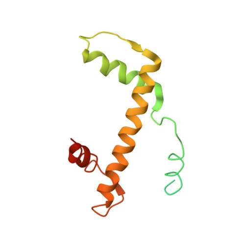
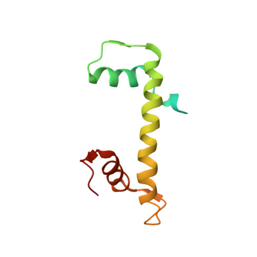
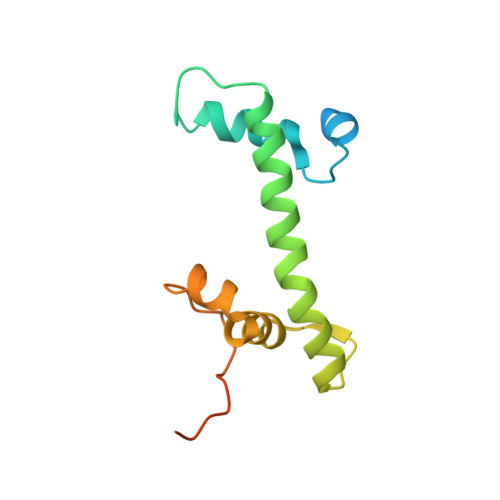
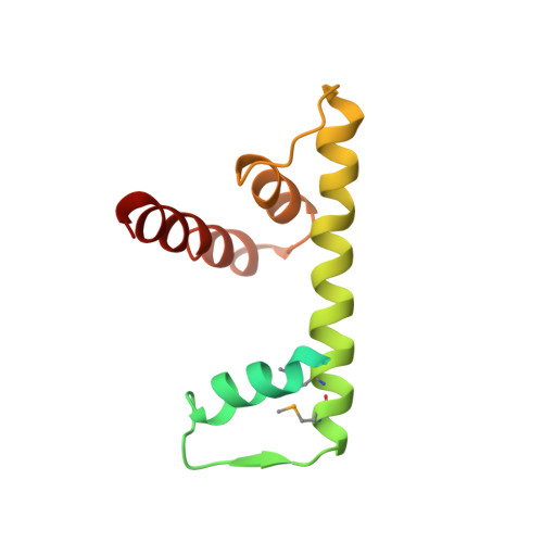
Crystal structure of the human centromeric nucleosome containing CENP-A

Tachiwana, H.Kagawa, W.Shiga, T.Osakabe, A.Miya, Y.Saito, K.Hayashi-Takanaka, Y.Oda, T.Sato, M.Park, S.-Y.Kimura, H.Kurumizaka, H.

(2011) Nature 476: 232-235

  • DOI: https://doi.org/10.1038/nature10258
  • Primary Citation of Related Structures:  
    3AN2

  • PubMed Abstract: 

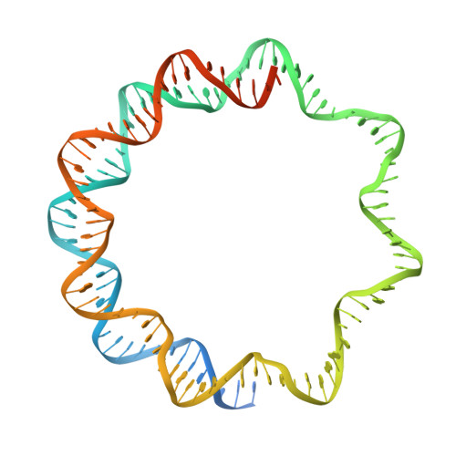
    In eukaryotes, accurate chromosome segregation during mitosis and meiosis is coordinated by kinetochores, which are unique chromosomal sites for microtubule attachment. Centromeres specify the kinetochore formation sites on individual chromosomes, and are epigenetically marked by the assembly of nucleosomes containing the centromere-specific histone H3 variant, CENP-A. Although the underlying mechanism is unclear, centromere inheritance is probably dictated by the architecture of the centromeric nucleosome. Here we report the crystal structure of the human centromeric nucleosome containing CENP-A and its cognate α-satellite DNA derivative (147 base pairs). In the human CENP-A nucleosome, the DNA is wrapped around the histone octamer, consisting of two each of histones H2A, H2B, H4 and CENP-A, in a left-handed orientation. However, unlike the canonical H3 nucleosome, only the central 121 base pairs of the DNA are visible. The thirteen base pairs from both ends of the DNA are invisible in the crystal structure, and the αN helix of CENP-A is shorter than that of H3, which is known to be important for the orientation of the DNA ends in the canonical H3 nucleosome. A structural comparison of the CENP-A and H3 nucleosomes revealed that CENP-A contains two extra amino acid residues (Arg 80 and Gly 81) in the loop 1 region, which is completely exposed to the solvent. Mutations of the CENP-A loop 1 residues reduced CENP-A retention at the centromeres in human cells. Therefore, the CENP-A loop 1 may function in stabilizing the centromeric chromatin containing CENP-A, possibly by providing a binding site for trans-acting factors. The structure provides the first atomic-resolution picture of the centromere-specific nucleosome.


  • Organizational Affiliation

    Laboratory of Structural Biology, Graduate School of Advanced Science and Engineering, Waseda University, 2-2 Wakamatsu-cho, Shinjuku-ku, Tokyo 162-8480, Japan.


Macromolecules

Find similar proteins by:  (by identity cutoff)  |  3D Structure
Entity ID: 1
MoleculeChains Sequence LengthOrganismDetailsImage
Histone H3-like centromeric protein A
A, E
143Homo sapiensMutation(s): 0 
Gene Names: CENPA
UniProt & NIH Common Fund Data Resources
Find proteins for P49450 (Homo sapiens)
Explore P49450 
Go to UniProtKB:  P49450
PHAROS:  P49450
GTEx:  ENSG00000115163 
Entity Groups  
Sequence Clusters30% Identity50% Identity70% Identity90% Identity95% Identity100% Identity
UniProt GroupP49450
Sequence Annotations
Expand
  • Reference Sequence
Find similar proteins by:  (by identity cutoff)  |  3D Structure
Entity ID: 2
MoleculeChains Sequence LengthOrganismDetailsImage
Histone H4
B, F
106Homo sapiensMutation(s): 0 
Gene Names: H4
UniProt & NIH Common Fund Data Resources
Find proteins for P62805 (Homo sapiens)
Explore P62805 
Go to UniProtKB:  P62805
PHAROS:  P62805
Entity Groups  
Sequence Clusters30% Identity50% Identity70% Identity90% Identity95% Identity100% Identity
UniProt GroupP62805
Sequence Annotations
Expand
  • Reference Sequence
Find similar proteins by:  (by identity cutoff)  |  3D Structure
Entity ID: 3
MoleculeChains Sequence LengthOrganismDetailsImage
Histone H2A type 1-B/E
C, G
133Homo sapiensMutation(s): 0 
Gene Names: H2A
UniProt & NIH Common Fund Data Resources
Find proteins for P04908 (Homo sapiens)
Explore P04908 
Go to UniProtKB:  P04908
PHAROS:  P04908
Entity Groups  
Sequence Clusters30% Identity50% Identity70% Identity90% Identity95% Identity100% Identity
UniProt GroupP04908
Sequence Annotations
Expand
  • Reference Sequence
Find similar proteins by:  (by identity cutoff)  |  3D Structure
Entity ID: 4
MoleculeChains Sequence LengthOrganismDetailsImage
Histone H2B type 1-J
D, H
129Homo sapiensMutation(s): 0 
Gene Names: H2B
UniProt & NIH Common Fund Data Resources
Find proteins for P06899 (Homo sapiens)
Explore P06899 
Go to UniProtKB:  P06899
PHAROS:  P06899
GTEx:  ENSG00000124635 
Entity Groups  
Sequence Clusters30% Identity50% Identity70% Identity90% Identity95% Identity100% Identity
UniProt GroupP06899
Sequence Annotations
Expand
  • Reference Sequence
Find similar nucleic acids by:  (by identity cutoff)  |  3D Structure
Entity ID: 5
MoleculeChains LengthOrganismImage
147 mer DNA
I, J
147N/A
Sequence Annotations
Expand
  • Reference Sequence
Small Molecules
Modified Residues  1 Unique
IDChains TypeFormula2D DiagramParent
MSE
Query on MSE
D, H
L-PEPTIDE LINKINGC5 H11 N O2 SeMET
Experimental Data & Validation

Experimental Data

  • Method: X-RAY DIFFRACTION
  • Resolution: 3.60 Å
  • R-Value Free: 0.325 
  • R-Value Work: 0.271 
  • R-Value Observed: 0.271 
  • Space Group: P 1 21 1
Unit Cell:
Length ( Å )Angle ( ˚ )
a = 65.84α = 90
b = 83.29β = 100.7
c = 176.83γ = 90
Software Package:
Software NamePurpose
BSSdata collection
MOLREPphasing
CNSrefinement
HKL-2000data reduction
HKL-2000data scaling

Structure Validation

View Full Validation Report


Entry History 

Deposition Data

Revision History  (Full details and data files)

  • Version 1.0: 2011-07-20
    Type: Initial release
  • Version 1.1: 2011-08-10
    Changes: Database references
  • Version 1.2: 2011-08-17
    Changes: Database references
  • Version 1.3: 2012-07-25
    Changes: Structure summary
  • Version 1.4: 2023-11-01
    Changes: Data collection, Database references, Derived calculations, Refinement description
  • Version 1.5: 2023-11-15
    Changes: Data collection