3E3D

Structure of hen egg white lysozyme with the magic triangle I3C


Experimental Data Snapshot

  • Method: X-RAY DIFFRACTION
  • Resolution: 1.55 Å
  • R-Value Free: 0.220 
  • R-Value Work: 0.171 
  • R-Value Observed: 0.173 

wwPDB Validation   3D Report Full Report


Ligand Structure Quality Assessment 


This is version 1.3 of the entry. See complete history


Literature

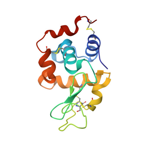
A magic triangle for experimental phasing of macromolecules

Beck, T.Krasauskas, A.Gruene, T.Sheldrick, G.M.

(2008) Acta Crystallogr D Biol Crystallogr 64: 1179-1182

  • DOI: https://doi.org/10.1107/S0907444908030266
  • Primary Citation of Related Structures:  
    3E3D, 3E3S, 3E3T

  • PubMed Abstract: 

    Obtaining phase information for the solution of macromolecular structures is still one of the bottlenecks in X-ray crystallography. 5-Amino-2,4,6-triiodoisophthalic acid (I3C), in which three covalently bound iodines form an equilateral triangle, was incorporated into proteins in order to obtain phases by single-wavelength anomalous dispersion (SAD). An improved binding capability compared with simple heavy-metal ions, ready availability, improved recognition of potential heavy-atom sites and low toxicity make I3C particularly suitable for experimental phasing.


  • Organizational Affiliation

    Department of Structural Chemistry, Georg-August-Universität Göttingen, Tammannstrasse 4, 37077 Göttingen, Germany. tbeck@shelx.uni-ac.gwdg.de


Macromolecules
Find similar proteins by:  (by identity cutoff)  |  3D Structure
Entity ID: 1
MoleculeChains Sequence LengthOrganismDetailsImage
Lysozyme C129Gallus gallusMutation(s): 0 
EC: 3.2.1.17
UniProt
Find proteins for P00698 (Gallus gallus)
Explore P00698 
Go to UniProtKB:  P00698
Entity Groups  
Sequence Clusters30% Identity50% Identity70% Identity90% Identity95% Identity100% Identity
UniProt GroupP00698
Sequence Annotations
Expand
  • Reference Sequence
Experimental Data & Validation

Experimental Data

  • Method: X-RAY DIFFRACTION
  • Resolution: 1.55 Å
  • R-Value Free: 0.220 
  • R-Value Work: 0.171 
  • R-Value Observed: 0.173 
  • Space Group: P 43 21 2
Unit Cell:
Length ( Å )Angle ( ˚ )
a = 76.83α = 90
b = 76.83β = 90
c = 38.87γ = 90
Software Package:
Software NamePurpose
SHELXrefinement
PDB_EXTRACTdata extraction
MAR345data collection
XDSdata reduction
SADABSdata scaling
SHELXDphasing
SHELXL-97refinement

Structure Validation

View Full Validation Report


Ligand Structure Quality Assessment 


Entry History 

Deposition Data

Revision History  (Full details and data files)

  • Version 1.0: 2008-10-28
    Type: Initial release
  • Version 1.1: 2011-07-13
    Changes: Version format compliance
  • Version 1.2: 2012-04-11
    Changes: Database references
  • Version 1.3: 2017-10-25
    Changes: Refinement description