3GJ1

Non photoactivated state of PA-GFP


Experimental Data Snapshot

  • Method: X-RAY DIFFRACTION
  • Resolution: 1.80 Å
  • R-Value Free: 0.252 
  • R-Value Work: 0.209 
  • R-Value Observed: 0.211 

wwPDB Validation   3D Report Full Report


This is version 1.5 of the entry. See complete history


Literature

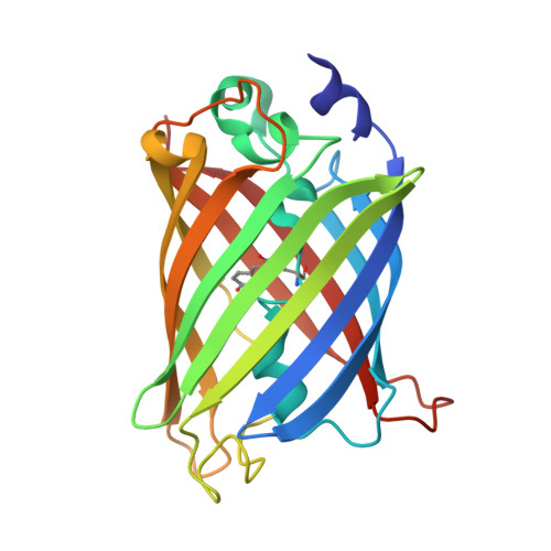
Structure and mechanism of the photoactivatable green fluorescent protein.

Henderson, J.N.Gepshtein, R.Heenan, J.R.Kallio, K.Huppert, D.Remington, S.J.

(2009) J Am Chem Soc 131: 4176-4177

  • DOI: https://doi.org/10.1021/ja808851n
  • Primary Citation of Related Structures:  
    3GJ1, 3GJ2

  • PubMed Abstract: 

    Crystal structures of the photoactivatable green fluorescent protein T203H variant (PA-GFP) have been solved in the native and photoactivated states, which under 488 nm illumination are dark and brightly fluorescent, respectively. We demonstrate that photoactivation of PA-GFP is the result of a UV-induced decarboxylation of the Glu222 side chain that shifts the chromophore equilibrium to the anionic form. Coupled with the T203H mutation, which stabilizes the native PA-GFP neutral chromophore, Glu222 decarboxylation yields a 100-fold contrast enhancement relative to wild-type GFP (WT). Additionally, the structures provide insights into the spectroscopic differences between WT and PA-GFP steady-state fluorescence maxima and excited-state proton transfer dynamics.


  • Organizational Affiliation

    Institute of Molecular Biology and Department of Physics, University of Oregon, Eugene, Oregon 97403-1229, USA.


Macromolecules
Find similar proteins by:  (by identity cutoff)  |  3D Structure
Entity ID: 1
MoleculeChains Sequence LengthOrganismDetailsImage
Green fluorescent protein
A, B, C, D
229Aequorea victoriaMutation(s): 5 
Gene Names: GFP
UniProt
Find proteins for P42212 (Aequorea victoria)
Explore P42212 
Go to UniProtKB:  P42212
Entity Groups  
Sequence Clusters30% Identity50% Identity70% Identity90% Identity95% Identity100% Identity
UniProt GroupP42212
Sequence Annotations
Expand
  • Reference Sequence
Small Molecules
Ligands 2 Unique
IDChains Name / Formula / InChI Key2D Diagram3D Interactions
SO4
Query on SO4

Download Ideal Coordinates CCD File 
E [auth A],
G [auth B],
J [auth C],
L [auth D],
M [auth D]
SULFATE ION
O4 S
QAOWNCQODCNURD-UHFFFAOYSA-L
CL
Query on CL

Download Ideal Coordinates CCD File 
F [auth A],
H [auth B],
I [auth B],
K [auth C],
N [auth D]
CHLORIDE ION
Cl
VEXZGXHMUGYJMC-UHFFFAOYSA-M
Modified Residues  1 Unique
IDChains TypeFormula2D DiagramParent
CRO
Query on CRO
A, B, C, D
L-PEPTIDE LINKINGC15 H17 N3 O5THR, TYR, GLY
Experimental Data & Validation

Experimental Data

  • Method: X-RAY DIFFRACTION
  • Resolution: 1.80 Å
  • R-Value Free: 0.252 
  • R-Value Work: 0.209 
  • R-Value Observed: 0.211 
  • Space Group: P 21 21 21
Unit Cell:
Length ( Å )Angle ( ˚ )
a = 84.515α = 90
b = 86.721β = 90
c = 144.159γ = 90
Software Package:
Software NamePurpose
DENZOdata reduction
SCALEPACKdata scaling
EPMRphasing
REFMACrefinement
PDB_EXTRACTdata extraction
HKL-2000data collection

Structure Validation

View Full Validation Report


Entry History 

Deposition Data

Revision History  (Full details and data files)

  • Version 1.0: 2009-03-24
    Type: Initial release
  • Version 1.1: 2011-07-13
    Changes: Version format compliance
  • Version 1.2: 2017-11-01
    Changes: Advisory, Refinement description
  • Version 1.3: 2021-10-20
    Changes: Database references, Derived calculations
  • Version 1.4: 2023-09-06
    Changes: Data collection, Refinement description
  • Version 1.5: 2023-11-22
    Changes: Data collection