3J46

Structure of the SecY protein translocation channel in action


Experimental Data Snapshot

  • Method: ELECTRON MICROSCOPY
  • Resolution: 10.1 Å
  • Aggregation State: PARTICLE 
  • Reconstruction Method: SINGLE PARTICLE 

wwPDB Validation   3D Report Full Report


This is version 2.0 of the entry. See complete history


Literature

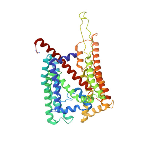
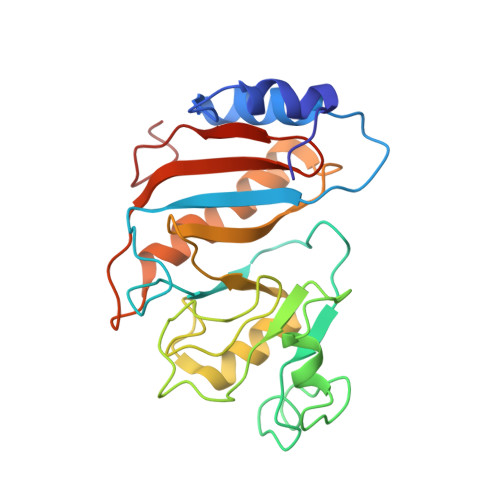
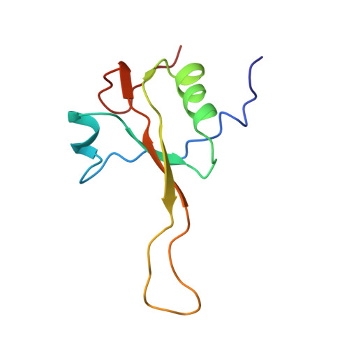
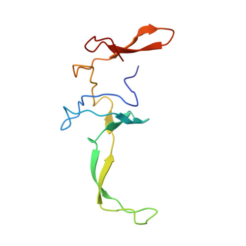
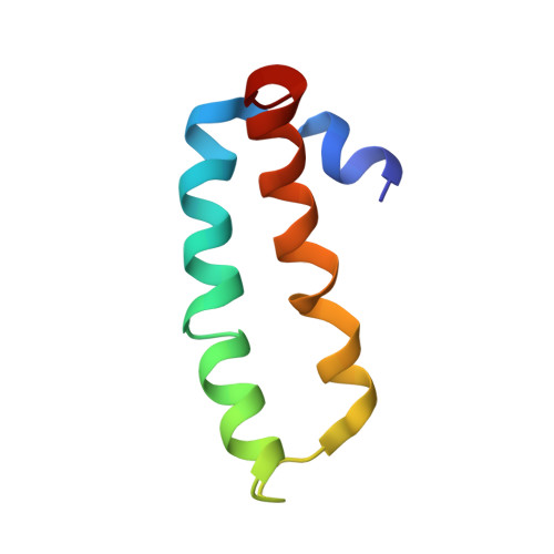
Structure of the SecY channel during initiation of protein translocation.

Park, E.Menetret, J.F.Gumbart, J.C.Ludtke, S.J.Li, W.Whynot, A.Rapoport, T.A.Akey, C.W.

(2013) Nature 506: 102-106

  • DOI: https://doi.org/10.1038/nature12720
  • Primary Citation of Related Structures:  
    3J45, 3J46, 4V4N

  • PubMed Abstract: 

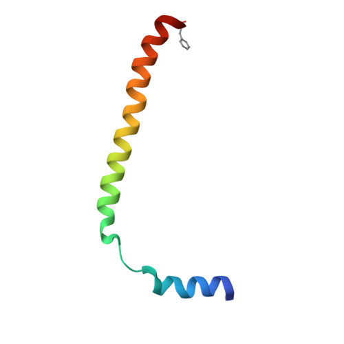
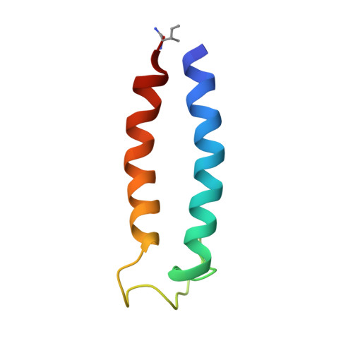
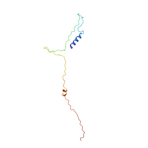
    Many secretory proteins are targeted by signal sequences to a protein-conducting channel, formed by prokaryotic SecY or eukaryotic Sec61 complexes, and are translocated across the membrane during their synthesis. Crystal structures of the inactive channel show that the SecY subunit of the heterotrimeric complex consists of two halves that form an hourglass-shaped pore with a constriction in the middle of the membrane and a lateral gate that faces the lipid phase. The closed channel has an empty cytoplasmic funnel and an extracellular funnel that is filled with a small helical domain, called the plug. During initiation of translocation, a ribosome-nascent chain complex binds to the SecY (or Sec61) complex, resulting in insertion of the nascent chain. However, the mechanism of channel opening during translocation is unclear. Here we have addressed this question by determining structures of inactive and active ribosome-channel complexes with cryo-electron microscopy. Non-translating ribosome-SecY channel complexes derived from Methanocaldococcus jannaschii or Escherichia coli show the channel in its closed state, and indicate that ribosome binding per se causes only minor changes. The structure of an active E. coli ribosome-channel complex demonstrates that the nascent chain opens the channel, causing mostly rigid body movements of the amino- and carboxy-terminal halves of SecY. In this early translocation intermediate, the polypeptide inserts as a loop into the SecY channel with the hydrophobic signal sequence intercalated into the open lateral gate. The nascent chain also forms a loop on the cytoplasmic surface of SecY rather than entering the channel directly.


  • Organizational Affiliation

    Department of Cell Biology and Howard Hughes Medical Institute, Harvard Medical School, 240 Longwood Avenue, Boston, Massachusetts 02115, USA.


Macromolecules

Find similar proteins by:  (by identity cutoff)  |  3D Structure
Entity ID: 1
MoleculeChains Sequence LengthOrganismDetailsImage
Protein translocase subunit SecYA [auth y]437Escherichia coliMutation(s): 3 
Membrane Entity: Yes 
UniProt
Find proteins for P0AGA2 (Escherichia coli (strain K12))
Explore P0AGA2 
Go to UniProtKB:  P0AGA2
Entity Groups  
Sequence Clusters30% Identity50% Identity70% Identity90% Identity95% Identity100% Identity
UniProt GroupP0AGA2
Sequence Annotations
Expand
  • Reference Sequence
Find similar proteins by:  (by identity cutoff)  |  3D Structure
Entity ID: 2
MoleculeChains Sequence LengthOrganismDetailsImage
Preprotein translocase subunit SecEB [auth E]56Escherichia coliMutation(s): 2 
Membrane Entity: Yes 
UniProt
Find proteins for P0AG96 (Escherichia coli (strain K12))
Explore P0AG96 
Go to UniProtKB:  P0AG96
Entity Groups  
Sequence Clusters30% Identity50% Identity70% Identity90% Identity95% Identity100% Identity
UniProt GroupP0AG96
Sequence Annotations
Expand
  • Reference Sequence
Find similar proteins by:  (by identity cutoff)  |  3D Structure
Entity ID: 3
MoleculeChains Sequence LengthOrganismDetailsImage
Protein-export membrane protein SecGC [auth G]67Escherichia coliMutation(s): 2 
Membrane Entity: Yes 
UniProt
Find proteins for P0AG99 (Escherichia coli (strain K12))
Explore P0AG99 
Go to UniProtKB:  P0AG99
Entity Groups  
Sequence Clusters30% Identity50% Identity70% Identity90% Identity95% Identity100% Identity
UniProt GroupP0AG99
Sequence Annotations
Expand
  • Reference Sequence
Find similar proteins by:  (by identity cutoff)  |  3D Structure
Entity ID: 4
MoleculeChains Sequence LengthOrganismDetailsImage
NC100D [auth n]101Escherichia coliMutation(s): 0 
Entity Groups  
Sequence Clusters30% Identity50% Identity70% Identity90% Identity95% Identity100% Identity
Sequence Annotations
Expand
  • Reference Sequence
Find similar proteins by:  (by identity cutoff)  |  3D Structure
Entity ID: 7
MoleculeChains Sequence LengthOrganismDetailsImage
50S ribosomal protein L1G [auth 5]234Escherichia coliMutation(s): 0 
UniProt
Find proteins for P0A7L0 (Escherichia coli (strain K12))
Explore P0A7L0 
Go to UniProtKB:  P0A7L0
Entity Groups  
Sequence Clusters30% Identity50% Identity70% Identity90% Identity95% Identity100% Identity
UniProt GroupP0A7L0
Sequence Annotations
Expand
  • Reference Sequence
Find similar proteins by:  (by identity cutoff)  |  3D Structure
Entity ID: 8
MoleculeChains Sequence LengthOrganismDetailsImage
50S ribosomal protein L23PH [auth T]100Escherichia coliMutation(s): 0 
UniProt
Find proteins for P0ADZ0 (Escherichia coli (strain K12))
Explore P0ADZ0 
Go to UniProtKB:  P0ADZ0
Entity Groups  
Sequence Clusters30% Identity50% Identity70% Identity90% Identity95% Identity100% Identity
UniProt GroupP0ADZ0
Sequence Annotations
Expand
  • Reference Sequence
Find similar proteins by:  (by identity cutoff)  |  3D Structure
Entity ID: 9
MoleculeChains Sequence LengthOrganismDetailsImage
50S ribosomal protein L24PI [auth U]103Escherichia coliMutation(s): 0 
UniProt
Find proteins for P60624 (Escherichia coli (strain K12))
Explore P60624 
Go to UniProtKB:  P60624
Entity Groups  
Sequence Clusters30% Identity50% Identity70% Identity90% Identity95% Identity100% Identity
UniProt GroupP60624
Sequence Annotations
Expand
  • Reference Sequence
Find similar proteins by:  (by identity cutoff)  |  3D Structure
Entity ID: 10
MoleculeChains Sequence LengthOrganismDetailsImage
50S ribosomal protein L29PJ [auth Y]63Escherichia coliMutation(s): 0 
UniProt
Find proteins for P0A7M6 (Escherichia coli (strain K12))
Explore P0A7M6 
Go to UniProtKB:  P0A7M6
Entity Groups  
Sequence Clusters30% Identity50% Identity70% Identity90% Identity95% Identity100% Identity
UniProt GroupP0A7M6
Sequence Annotations
Expand
  • Reference Sequence
Find similar nucleic acids by:  (by identity cutoff)  |  3D Structure
Entity ID: 5
MoleculeChains LengthOrganismImage
P-tRNAE [auth p]76Escherichia coli
Sequence Annotations
Expand
  • Reference Sequence
Find similar nucleic acids by:  (by identity cutoff)  |  3D Structure
Entity ID: 6
MoleculeChains LengthOrganismImage
A-tRNAF [auth a]76Escherichia coli
Sequence Annotations
Expand
  • Reference Sequence
Find similar nucleic acids by:  (by identity cutoff)  |  3D Structure
Entity ID: 11
MoleculeChains LengthOrganismImage
23S ribosomal RNAK [auth 1]63Escherichia coli
Sequence Annotations
Expand
  • Reference Sequence
Find similar nucleic acids by:  (by identity cutoff)  |  3D Structure
Entity ID: 12
MoleculeChains LengthOrganismImage
23S ribosomal RNAL [auth 2]36Escherichia coli
Sequence Annotations
Expand
  • Reference Sequence
Find similar nucleic acids by:  (by identity cutoff)  |  3D Structure
Entity ID: 13
MoleculeChains LengthOrganismImage
23S ribosomal RNAM [auth 3]44Escherichia coli
Sequence Annotations
Expand
  • Reference Sequence
Find similar nucleic acids by:  (by identity cutoff)  |  3D Structure
Entity ID: 14
MoleculeChains LengthOrganismImage
23S ribosomal RNAN [auth 4]109Escherichia coli
Sequence Annotations
Expand
  • Reference Sequence
Experimental Data & Validation

Experimental Data

  • Method: ELECTRON MICROSCOPY
  • Resolution: 10.1 Å
  • Aggregation State: PARTICLE 
  • Reconstruction Method: SINGLE PARTICLE 
EM Software:
TaskSoftware PackageVersion
RECONSTRUCTIONEMAN2

Structure Validation

View Full Validation Report


Entry History 

Deposition Data

Revision History  (Full details and data files)

  • Version 1.0: 2013-10-23
    Type: Initial release
  • Version 1.1: 2013-11-06
    Changes: Database references
  • Version 1.2: 2014-02-05
    Changes: Database references
  • Version 1.3: 2018-07-18
    Changes: Data collection
  • Version 2.0: 2019-07-03
    Changes: Data collection, Derived calculations, Non-polymer description, Structure summary