3JYT

K65R mutant HIV-1 reverse transcriptase cross-linked to DS-DNA and complexed with DATP as the incoming nucleotide substrate


Experimental Data Snapshot

  • Method: X-RAY DIFFRACTION
  • Resolution: 3.30 Å
  • R-Value Free: 0.287 
  • R-Value Work: 0.253 
  • R-Value Observed: 0.253 

wwPDB Validation   3D Report Full Report


Ligand Structure Quality Assessment 


This is version 2.1 of the entry. See complete history


Literature

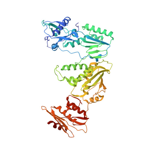
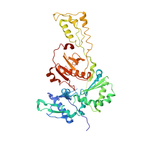
Structural basis for the role of the K65r mutation in HIV-1 reverse transcriptase polymerization, excision antagonism, and tenofovir resistance.

Das, K.Bandwar, R.P.White, K.L.Feng, J.Y.Sarafianos, S.G.Tuske, S.Tu, X.Clark, A.D.Boyer, P.L.Hou, X.Gaffney, B.L.Jones, R.A.Miller, M.D.Hughes, S.H.Arnold, E.

(2009) J Biol Chem 284: 35092-35100

  • DOI: https://doi.org/10.1074/jbc.M109.022525
  • Primary Citation of Related Structures:  
    3JSM, 3JYT

  • PubMed Abstract: 

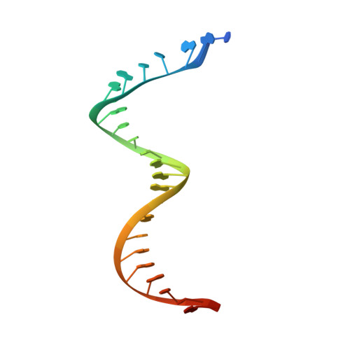
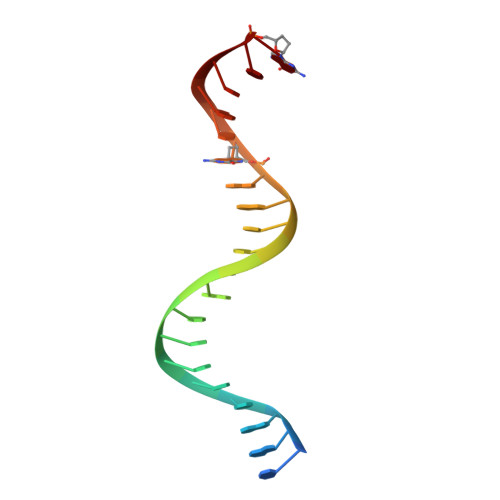
    K65R is a primary reverse transcriptase (RT) mutation selected in human immunodeficiency virus type 1-infected patients taking antiretroviral regimens containing tenofovir disoproxil fumarate or other nucleoside analog RT drugs. We determined the crystal structures of K65R mutant RT cross-linked to double-stranded DNA and in complexes with tenofovir diphosphate (TFV-DP) or dATP. The crystals permit substitution of TFV-DP with dATP at the dNTP-binding site. The guanidinium planes of the arginines K65R and Arg(72) were stacked to form a molecular platform that restricts the conformational adaptability of both of the residues, which explains the negative effects of the K65R mutation on nucleotide incorporation and on excision. Furthermore, the guanidinium planes of K65R and Arg(72) were stacked in two different rotameric conformations in TFV-DP- and dATP-bound structures that may help explain how K65R RT discriminates the drug from substrates. These K65R-mediated effects on RT structure and function help us to visualize the complex interaction with other key nucleotide RT drug resistance mutations, such as M184V, L74V, and thymidine analog resistance mutations.


  • Organizational Affiliation

    Center for Advanced Biotechnology and Medicine (CABM), Rutgers University, Piscataway, New Jersey 08854, USA.


Macromolecules

Find similar proteins by:  (by identity cutoff)  |  3D Structure
Entity ID: 1
MoleculeChains Sequence LengthOrganismDetailsImage
Reverse transcriptase/ribonuclease H558Human immunodeficiency virus type 1 BH10Mutation(s): 3 
Gene Names: gag-pol
EC: 2.7.7.49
UniProt
Find proteins for P03366 (Human immunodeficiency virus type 1 group M subtype B (isolate BH10))
Explore P03366 
Go to UniProtKB:  P03366
Entity Groups  
Sequence Clusters30% Identity50% Identity70% Identity90% Identity95% Identity100% Identity
UniProt GroupP03366
Sequence Annotations
Expand
  • Reference Sequence
Find similar proteins by:  (by identity cutoff)  |  3D Structure
Entity ID: 2
MoleculeChains Sequence LengthOrganismDetailsImage
p51 RT437Human immunodeficiency virus type 1 BH10Mutation(s): 1 
Gene Names: gag-pol
UniProt
Find proteins for P03366 (Human immunodeficiency virus type 1 group M subtype B (isolate BH10))
Explore P03366 
Go to UniProtKB:  P03366
Entity Groups  
Sequence Clusters30% Identity50% Identity70% Identity90% Identity95% Identity100% Identity
UniProt GroupP03366
Sequence Annotations
Expand
  • Reference Sequence
Find similar nucleic acids by:  (by identity cutoff)  |  3D Structure
Entity ID: 3
MoleculeChains LengthOrganismImage
DNA (5'-D(*A*TP*GP*GP*TP*CP*GP*GP*CP*GP*CP*CP*CP*GP*AP*AP*CP*AP*GP*GP*GP*AP*CP*TP*GP*TP*G)-3')C [auth T]27synthetic construct
Sequence Annotations
Expand
  • Reference Sequence

Find similar nucleic acids by:  Sequence   |   3D Structure  

Entity ID: 4
MoleculeChains LengthOrganismImage
DNA (5'-D(*A*CP*AP*GP*TP*CP*CP*CP*TP*GP*TP*TP*CP*GP*GP*(MRG)P*CP*GP*CP*CP*(DDG))-3')D [auth P]21synthetic construct
Sequence Annotations
Expand
  • Reference Sequence
Experimental Data & Validation

Experimental Data

  • Method: X-RAY DIFFRACTION
  • Resolution: 3.30 Å
  • R-Value Free: 0.287 
  • R-Value Work: 0.253 
  • R-Value Observed: 0.253 
  • Space Group: P 31 1 2
Unit Cell:
Length ( Å )Angle ( ˚ )
a = 169.72α = 90
b = 169.72β = 90
c = 155.41γ = 120
Software Package:
Software NamePurpose
ADSCdata collection
CNSrefinement
HKL-2000data reduction
SCALEPACKdata scaling
CNSphasing

Structure Validation

View Full Validation Report


Ligand Structure Quality Assessment 


Entry History 

Deposition Data

Revision History  (Full details and data files)

  • Version 1.0: 2009-10-06
    Type: Initial release
  • Version 1.1: 2011-07-13
    Changes: Version format compliance
  • Version 2.0: 2021-10-13
    Changes: Advisory, Atomic model, Data collection, Database references, Derived calculations, Source and taxonomy, Structure summary
  • Version 2.1: 2023-09-06
    Changes: Data collection, Refinement description