3KRD

Crystal Structure of Mycobacterium Tuberculosis Proteasome in complex with Fellutamide B


Experimental Data Snapshot

  • Method: X-RAY DIFFRACTION
  • Resolution: 2.50 Å
  • R-Value Free: 0.229 
  • R-Value Work: 0.208 
  • R-Value Observed: 0.208 

wwPDB Validation   3D Report Full Report


Ligand Structure Quality Assessment 


This is version 2.0 of the entry. See complete history


Literature

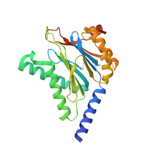
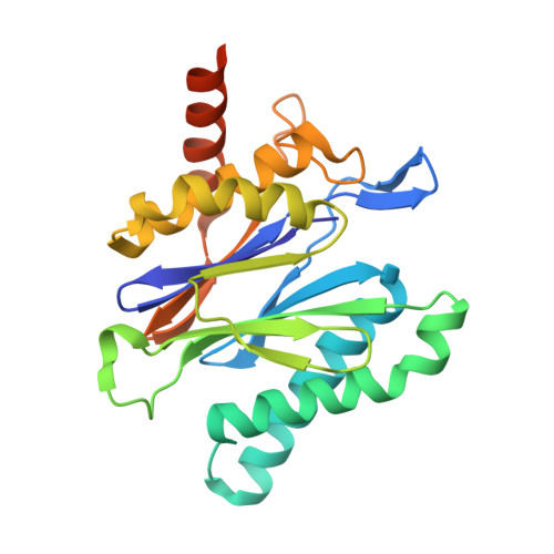
Fellutamide B is a potent inhibitor of the Mycobacterium tuberculosis proteasome.

Lin, G.Li, D.Chidawanyika, T.Nathan, C.Li, H.

(2010) Arch Biochem Biophys 501: 214-220

  • DOI: https://doi.org/10.1016/j.abb.2010.06.009
  • Primary Citation of Related Structures:  
    3KRD

  • PubMed Abstract: 

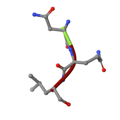
    Via high-throughput screening of a natural compound library, we have identified a lipopeptide aldehyde, fellutamide B (1), as the most potent inhibitor of the Mycobacterium tuberculosis (Mtb) proteasome tested to date. Kinetic studies reveal that 1 inhibits both Mtb and human proteasomes in a time-dependent manner under steady-state condition. Remarkably, 1 inhibits the Mtb proteasome in a single-step binding mechanism with K(i)=6.8 nM, whereas it inhibits the human proteasome beta5 active site following a two-step mechanism with K(i)=11.5 nM and K(i)(*)=0.93 nM. Co-crystallization of 1 bound to the Mtb proteasome revealed a structural basis for the tight binding of 1 to the active sites of the Mtb proteasome. The hemiacetal group of 1 in the Mtb proteasome takes the (R)-configuration, whereas in the yeast proteasome it takes the (S)-configuration, indicating that the pre-chiral CHO group of 1 binds to the active site Thr1 in a different orientation. Re-examination of the structure of the yeast proteasome in complex with 1 showed significant conformational changes at the substrate-binding cleft along the active site. These structural differences are consistent with the different kinetic mechanisms of 1 against Mtb and human proteasomes.


  • Organizational Affiliation

    Department of Microbiology and Immunology, Weill Medical College of Cornell University, 1300 York Ave., New York, NY 10065, United States. gal2005@med.cornell.edu


Macromolecules
Find similar proteins by:  (by identity cutoff)  |  3D Structure
Entity ID: 1
MoleculeChains Sequence LengthOrganismDetailsImage
Proteasome subunit alpha
A,
AA [auth 1],
B,
D,
F,
I,
K,
M,
O,
Q,
S,
U,
W,
Y
248Mycobacterium tuberculosisMutation(s): 0 
Gene Names: prcAMRA_2124
UniProt
Find proteins for P9WHU1 (Mycobacterium tuberculosis (strain ATCC 25618 / H37Rv))
Explore P9WHU1 
Go to UniProtKB:  P9WHU1
Entity Groups  
Sequence Clusters30% Identity50% Identity70% Identity90% Identity95% Identity100% Identity
UniProt GroupP9WHU1
Sequence Annotations
Expand
  • Reference Sequence
Find similar proteins by:  (by identity cutoff)  |  3D Structure
Entity ID: 2
MoleculeChains Sequence LengthOrganismDetailsImage
Proteasome subunit beta
BA [auth 2],
C,
E,
G,
H,
J,
L,
N,
P,
R,
T,
V,
X,
Z
240Mycobacterium tuberculosisMutation(s): 0 
Gene Names: prcBMRA_2125
EC: 3.4.25.1
UniProt
Find proteins for P9WHT9 (Mycobacterium tuberculosis (strain ATCC 25618 / H37Rv))
Explore P9WHT9 
Go to UniProtKB:  P9WHT9
Entity Groups  
Sequence Clusters30% Identity50% Identity70% Identity90% Identity95% Identity100% Identity
UniProt GroupP9WHT9
Sequence Annotations
Expand
  • Reference Sequence

Find similar proteins by:  Sequence   |   3D Structure  

Entity ID: 3
MoleculeChains Sequence LengthOrganismDetailsImage
Fellutamide B3synthetic constructMutation(s): 0 
Sequence Annotations
Expand
  • Reference Sequence
Small Molecules
Ligands 1 Unique
IDChains Name / Formula / InChI Key2D Diagram3D Interactions
HXD
Query on HXD

Download Ideal Coordinates CCD File 
AB [auth k]
BB [auth l]
CB [auth m]
DB [auth n]
QA [auth a]
AB [auth k],
BB [auth l],
CB [auth m],
DB [auth n],
QA [auth a],
RA [auth b],
SA [auth c],
TA [auth d],
UA [auth e],
VA [auth f],
WA [auth g],
XA [auth h],
YA [auth i],
ZA [auth j]
(3R)-3-HYDROXYDODECANOIC ACID
C12 H24 O3
MUCMKTPAZLSKTL-LLVKDONJSA-N
Binding Affinity Annotations 
IDSourceBinding Affinity
FEB PDBBind:  3KRD Ki: 6.8 (nM) from 1 assay(s)
Experimental Data & Validation

Experimental Data

  • Method: X-RAY DIFFRACTION
  • Resolution: 2.50 Å
  • R-Value Free: 0.229 
  • R-Value Work: 0.208 
  • R-Value Observed: 0.208 
  • Space Group: P 1 21 1
Unit Cell:
Length ( Å )Angle ( ˚ )
a = 170.186α = 90
b = 118.099β = 112.62
c = 194.347γ = 90
Software Package:
Software NamePurpose
CBASSdata collection
CNSrefinement
HKL-2000data reduction
HKL-2000data scaling
CNSphasing

Structure Validation

View Full Validation Report


Ligand Structure Quality Assessment 


Entry History 

Deposition Data

  • Released Date: 2010-09-29 
  • Deposition Author(s): Li, D., Li, H.

Revision History  (Full details and data files)

  • Version 1.0: 2010-09-29
    Type: Initial release
  • Version 1.1: 2011-07-13
    Changes: Version format compliance
  • Version 2.0: 2024-03-27
    Changes: Advisory, Atomic model, Data collection, Database references, Derived calculations, Non-polymer description, Polymer sequence, Source and taxonomy, Structure summary