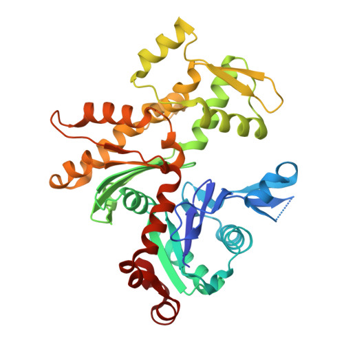
3M6G

Crystal structure of actin in complex with lobophorolide


Experimental Data Snapshot

  • Method: X-RAY DIFFRACTION
  • Resolution: 2.00 Å
  • R-Value Free: 0.227 
  • R-Value Work: 0.183 
  • R-Value Observed: 0.185 

wwPDB Validation   3D Report Full Report


Ligand Structure Quality Assessment 


This is version 1.5 of the entry. See complete history


Literature

Two molecules of lobophorolide cooperate to stabilize an actin dimer using both their "ring" and "tail" region.

Blain, J.C.Mok, Y.F.Kubanek, J.Allingham, J.S.

(2010) Chem Biol 17: 802-807

  • DOI: https://doi.org/10.1016/j.chembiol.2010.06.010
  • Primary Citation of Related Structures:  
    3M6G

  • PubMed Abstract: 

    Actin filament-disrupting marine macrolides are promising templates from which to design therapeutics against cancer and other diseases that co-opt the actin cytoskeleton. Typically, these macrolides form either a 1:1 or 2:1 actin-macrolide complex where their aliphatic side chain, or "tail," has been reported to convey the major determinant of cytotoxicity. We now report the structure of the marine macrolide lobophorolide bound to actin with a unique 2:2 stoichiometry in which two lobophorolide molecules cooperate to form a dimerization interface that is composed entirely of the macrolide "ring" region, and each molecule of lobophorolide interacts with both actin subunits via their ring and tail regions to tether the subunits together. This binding mode imposes multiple barriers against microfilament stability and holds important implications for development of actin-targeting drugs and the evolution of macrolide biosynthetic enzymes.


  • Organizational Affiliation

    Department of Biochemistry, Queen's University, Kingston, ON K7L 3N6, Canada.


Macromolecules
Find similar proteins by:  (by identity cutoff)  |  3D Structure
Entity ID: 1
MoleculeChains Sequence LengthOrganismDetailsImage
Actin, alpha skeletal muscle
A, B
371Oryctolagus cuniculusMutation(s): 0 
UniProt
Find proteins for P68135 (Oryctolagus cuniculus)
Explore P68135 
Go to UniProtKB:  P68135
Entity Groups  
Sequence Clusters30% Identity50% Identity70% Identity90% Identity95% Identity100% Identity
UniProt GroupP68135
Sequence Annotations
Expand
  • Reference Sequence
Small Molecules
Ligands 4 Unique
IDChains Name / Formula / InChI Key2D Diagram3D Interactions
LO3
Query on LO3

Download Ideal Coordinates CCD File 
E [auth A],
J [auth B]
(1S,3S,4S,5S,7R,8S,9R,12E,14E,16R,17R,19R)-16-hydroxy-9-{(1S,2S,3S)-2-hydroxy-5-[(2S,4R,6S)-4-methoxy-6-methyltetrahydro-2H-pyran-2-yl]-1,3-dimethylpentyl}-3,5,7,17-tetramethoxy-8,14-dimethyl-11H-spiro[10,23-dioxabicyclo[17.3.1]tricosa-12,14,20-triene-4,2'-oxiran]-11-one
C42 H70 O12
YPLVKLZVFQFTLN-NNZJXHHNSA-N
ATP
Query on ATP

Download Ideal Coordinates CCD File 
D [auth A],
I [auth B]
ADENOSINE-5'-TRIPHOSPHATE
C10 H16 N5 O13 P3
ZKHQWZAMYRWXGA-KQYNXXCUSA-N
CA
Query on CA

Download Ideal Coordinates CCD File 
F [auth A],
G [auth A],
K [auth B],
L [auth B]
CALCIUM ION
Ca
BHPQYMZQTOCNFJ-UHFFFAOYSA-N
MG
Query on MG

Download Ideal Coordinates CCD File 
C [auth A],
H [auth B]
MAGNESIUM ION
Mg
JLVVSXFLKOJNIY-UHFFFAOYSA-N
Experimental Data & Validation

Experimental Data

  • Method: X-RAY DIFFRACTION
  • Resolution: 2.00 Å
  • R-Value Free: 0.227 
  • R-Value Work: 0.183 
  • R-Value Observed: 0.185 
  • Space Group: P 1 21 1
Unit Cell:
Length ( Å )Angle ( ˚ )
a = 53.932α = 90
b = 86.419β = 94.91
c = 100.868γ = 90
Software Package:
Software NamePurpose
DENZOdata reduction
SCALEPACKdata scaling
PHASERphasing
REFMACrefinement
PDB_EXTRACTdata extraction
HKL-2000data collection

Structure Validation

View Full Validation Report


Ligand Structure Quality Assessment 


Entry History 

Deposition Data

Revision History  (Full details and data files)

  • Version 1.0: 2010-09-08
    Type: Initial release
  • Version 1.1: 2011-07-13
    Changes: Version format compliance
  • Version 1.2: 2015-01-28
    Changes: Database references
  • Version 1.3: 2017-11-08
    Changes: Refinement description
  • Version 1.4: 2023-09-06
    Changes: Data collection, Database references, Derived calculations, Refinement description
  • Version 1.5: 2024-03-13
    Changes: Source and taxonomy