3MN0

Introducing a 2-His-1-Glu Non-Heme Iron Center into Myoglobin confers Nitric Oxide Reductase activity: Cu(II)-CN-FeBMb(-His) form


Experimental Data Snapshot

  • Method: X-RAY DIFFRACTION
  • Resolution: 1.65 Å
  • R-Value Free: 0.273 
  • R-Value Work: 0.245 

wwPDB Validation   3D Report Full Report


Ligand Structure Quality Assessment 


This is version 1.3 of the entry. See complete history


Literature

Introducing a 2-his-1-glu nonheme iron center into myoglobin confers nitric oxide reductase activity.

Lin, Y.W.Yeung, N.Gao, Y.G.Miner, K.D.Lei, L.Robinson, H.Lu, Y.

(2010) J Am Chem Soc 132: 9970-9972

  • DOI: https://doi.org/10.1021/ja103516n
  • Primary Citation of Related Structures:  
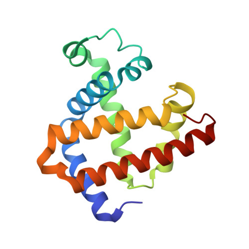
    3MN0

  • PubMed Abstract: 

    A conserved 2-His-1-Glu metal center, as found in natural nonheme iron-containing enzymes, was engineered into sperm whale myoglobin by replacing Leu29 and Phe43 with Glu and His, respectively (swMb L29E, F43H, H64, called Fe(B)Mb(-His)). A high resolution (1.65 A) crystal structure of Cu(II)-CN(-)-Fe(B)Mb(-His) was determined, demonstrating that the unique 2-His-1-Glu metal center was successfully created within swMb. The Fe(B)Mb(-His) can bind Cu, Fe, or Zn ions, with both Cu(I)-Fe(B)Mb(-His) and Fe(II)-Fe(B)Mb(-His) exhibiting nitric oxide reductase (NOR) activities. Cu dependent NOR activity was significantly higher than that of Fe in the same metal binding site. EPR studies showed that the reduction of NO to N(2)O catalyzed by these two enzymes resulted in different intermediates; a five-coordinate heme-NO species was observed for Cu(I)-Fe(B)Mb(-His) due to the cleavage of the proximal heme Fe-His bond, while Fe(II)-Fe(B)Mb(-His) remained six-coordinate. Therefore, both the metal ligand, Glu29, and the metal itself, Cu or Fe, play crucial roles in NOR activity. This study presents a novel protein model of NOR and provides insights into a newly discovered member of the NOR family, gNOR.


  • Organizational Affiliation

    Department of Chemistry, University of Illinois at Urbana-Champaign, Urbana, Illinois 61801, USA.


Macromolecules
Find similar proteins by:  (by identity cutoff)  |  3D Structure
Entity ID: 1
MoleculeChains Sequence LengthOrganismDetailsImage
Myoglobin153Physeter catodonMutation(s): 2 
Gene Names: MBpMbt7-7
UniProt
Find proteins for P02185 (Physeter macrocephalus)
Explore P02185 
Go to UniProtKB:  P02185
Entity Groups  
Sequence Clusters30% Identity50% Identity70% Identity90% Identity95% Identity100% Identity
UniProt GroupP02185
Sequence Annotations
Expand
  • Reference Sequence
Experimental Data & Validation

Experimental Data

  • Method: X-RAY DIFFRACTION
  • Resolution: 1.65 Å
  • R-Value Free: 0.273 
  • R-Value Work: 0.245 
  • Space Group: P 21 21 21
Unit Cell:
Length ( Å )Angle ( ˚ )
a = 39.61α = 90
b = 48.42β = 90
c = 77.63γ = 90
Software Package:
Software NamePurpose
HKL-2000data collection
SHELXmodel building
SHELXL-97refinement
MOLREPphasing
X-PLORrefinement
SHELXphasing

Structure Validation

View Full Validation Report


Ligand Structure Quality Assessment 


Entry History 

Deposition Data

Revision History  (Full details and data files)

  • Version 1.0: 2010-08-11
    Type: Initial release
  • Version 1.1: 2011-07-13
    Changes: Version format compliance
  • Version 1.2: 2021-10-06
    Changes: Database references, Derived calculations
  • Version 1.3: 2024-02-21
    Changes: Data collection