3PO6

Crystal structure of human carbonic anhydrase II with 6,7-Dimethoxy-1-methyl-3,4-dihydroisoquinoline-2(1H)-sulfonamide


Experimental Data Snapshot

  • Method: X-RAY DIFFRACTION
  • Resolution: 1.47 Å
  • R-Value Free: 0.175 
  • R-Value Work: 0.143 
  • R-Value Observed: 0.145 

wwPDB Validation   3D Report Full Report


Ligand Structure Quality Assessment 


This is version 1.3 of the entry. See complete history


Literature

Structural basis for the interaction between carbonic anhydrase and 1,2,3,4-tetrahydroisoquinolin-2-ylsulfonamides.

Mader, P.Brynda, J.Gitto, R.Agnello, S.Pachl, P.Supuran, C.T.Chimirri, A.Rezacova, P.

(2011) J Med Chem 54: 2522-2526

  • DOI: https://doi.org/10.1021/jm2000213
  • Primary Citation of Related Structures:  
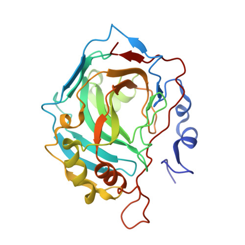
    3PO6

  • PubMed Abstract: 

    Isoquinolinesulfonamides inhibit human carbonic anhydrases (hCAs) and display selectivity toward therapeutically relevant isozymes. The crystal structure of hCA II in complex with 6,7-dimethoxy-1-methyl-1,2,3,4-tetrahydroisoquinolin-2-ylsulfonamide revealed unusual inhibitor binding. Structural analyses allowed for discerning the fine details of the inhibitor binding mode to the active site, thus providing clues for the future design of even more selective inhibitors for druggable isoforms such as the cancer associated hCA IX and neuronal hCA VII.


  • Organizational Affiliation

    Department of Structural Biology, Institute of Molecular Genetics, Academy of Sciences of the Czech Republic, Prague, Czech Republic.


Macromolecules
Find similar proteins by:  (by identity cutoff)  |  3D Structure
Entity ID: 1
MoleculeChains Sequence LengthOrganismDetailsImage
Carbonic anhydrase 2259Homo sapiensMutation(s): 0 
EC: 4.2.1.1
UniProt & NIH Common Fund Data Resources
Find proteins for P00918 (Homo sapiens)
Explore P00918 
Go to UniProtKB:  P00918
PHAROS:  P00918
GTEx:  ENSG00000104267 
Entity Groups  
Sequence Clusters30% Identity50% Identity70% Identity90% Identity95% Identity100% Identity
UniProt GroupP00918
Sequence Annotations
Expand
  • Reference Sequence
Small Molecules
Ligands 5 Unique
IDChains Name / Formula / InChI Key2D Diagram3D Interactions
RDT
Query on RDT

Download Ideal Coordinates CCD File 
D [auth A](1R)-6,7-dimethoxy-1-methyl-3,4-dihydroisoquinoline-2(1H)-sulfonamide
C12 H18 N2 O4 S
GUJQIHRSATWPQA-MRVPVSSYSA-N
DT9
Query on DT9

Download Ideal Coordinates CCD File 
C [auth A](1S)-6,7-dimethoxy-1-methyl-3,4-dihydroisoquinoline-2(1H)-sulfonamide
C12 H18 N2 O4 S
GUJQIHRSATWPQA-QMMMGPOBSA-N
GOL
Query on GOL

Download Ideal Coordinates CCD File 
E [auth A],
F [auth A],
G [auth A],
H [auth A]
GLYCEROL
C3 H8 O3
PEDCQBHIVMGVHV-UHFFFAOYSA-N
ZN
Query on ZN

Download Ideal Coordinates CCD File 
B [auth A]ZINC ION
Zn
PTFCDOFLOPIGGS-UHFFFAOYSA-N
ACT
Query on ACT

Download Ideal Coordinates CCD File 
I [auth A]ACETATE ION
C2 H3 O2
QTBSBXVTEAMEQO-UHFFFAOYSA-M
Binding Affinity Annotations 
IDSourceBinding Affinity
DT9 PDBBind:  3PO6 Ki: 87.3 (nM) from 1 assay(s)
ACT BindingDB:  3PO6 Ki: 1.30e+5 (nM) from 1 assay(s)
Experimental Data & Validation

Experimental Data

  • Method: X-RAY DIFFRACTION
  • Resolution: 1.47 Å
  • R-Value Free: 0.175 
  • R-Value Work: 0.143 
  • R-Value Observed: 0.145 
  • Space Group: P 1 21 1
Unit Cell:
Length ( Å )Angle ( ˚ )
a = 42.18α = 90
b = 41.21β = 104.31
c = 72γ = 90
Software Package:
Software NamePurpose
REFMACrefinement
MOSFLMdata reduction
SCALAdata scaling

Structure Validation

View Full Validation Report


Ligand Structure Quality Assessment 


Entry History 

Deposition Data

Revision History  (Full details and data files)

  • Version 1.0: 2011-04-06
    Type: Initial release
  • Version 1.1: 2011-07-13
    Changes: Version format compliance
  • Version 1.2: 2011-12-07
    Changes: Database references
  • Version 1.3: 2024-03-20
    Changes: Data collection, Database references, Derived calculations