3RN3

SEGMENTED ANISOTROPIC REFINEMENT OF BOVINE RIBONUCLEASE A BY THE APPLICATION OF THE RIGID-BODY TLS MODEL


Experimental Data Snapshot

  • Method: X-RAY DIFFRACTION
  • Resolution: 1.45 Å
  • R-Value Observed: 0.223 

wwPDB Validation   3D Report Full Report


This is version 1.3 of the entry. See complete history


Literature

Segmented anisotropic refinement of bovine ribonuclease A by the application of the rigid-body TLS model.

Howlin, B.Moss, D.S.Harris, G.W.

(1989) Acta Crystallogr A 45: 851-861

  • DOI: https://doi.org/10.1107/s0108767389009177
  • Primary Citation of Related Structures:  
    3RN3

  • PubMed Abstract: 

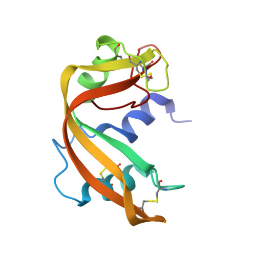
    The anisotropic displacements of selected rigid groups in bovine ribonuclease A have been refined from X-ray diffraction data by the application of the rigid-body TLS model. The rigid groups chosen were the side chains of tyrosine, histidine and phenylalanine and the planar side chains of aspartic acid, glutamic acid, glutamine, asparagine and arginine. The method has also been applied to the co-crystallizing active-site sulfate anion. This has enabled the description of the motion of the above-mentioned side-chain atoms by anisotropic displacement ellipsoids from a 1.45 A refinement. The hydrophobic side groups in the protein core show mainly translational motion, with mean-square librations of 20 deg2 which are similar to those found in some close-packed crystals of small organic molecules. Librational displacements are much more significant in the hydrophilic side groups where their magnitudes can be correlated with solvent accessibility. Large librations of some solvent exposed side chains correspond with the breakdown of a simple TLS model and the existence of multiple orientations of the side groups. The TLS model has also been applied to the whole protein molecule and shows that the average motion is approximately isotropic with little librational character.


  • Organizational Affiliation

    Department of Crystallography, Birkbeck College, London, England.


Macromolecules
Find similar proteins by:  (by identity cutoff)  |  3D Structure
Entity ID: 1
MoleculeChains Sequence LengthOrganismDetailsImage
RIBONUCLEASE A124Bos taurusMutation(s): 0 
EC: 3.1.27.5
UniProt
Find proteins for P61823 (Bos taurus)
Explore P61823 
Go to UniProtKB:  P61823
Entity Groups  
Sequence Clusters30% Identity50% Identity70% Identity90% Identity95% Identity100% Identity
UniProt GroupP61823
Sequence Annotations
Expand
  • Reference Sequence
Small Molecules
Ligands 1 Unique
IDChains Name / Formula / InChI Key2D Diagram3D Interactions
SO4
Query on SO4

Download Ideal Coordinates CCD File 
B [auth A]SULFATE ION
O4 S
QAOWNCQODCNURD-UHFFFAOYSA-L
Experimental Data & Validation

Experimental Data

  • Method: X-RAY DIFFRACTION
  • Resolution: 1.45 Å
  • R-Value Observed: 0.223 
  • Space Group: P 1 21 1
Unit Cell:
Length ( Å )Angle ( ˚ )
a = 30.45α = 90
b = 38.369β = 105.96
c = 53.215γ = 90
Software Package:
Software NamePurpose
RESTRAINrefinement

Structure Validation

View Full Validation Report


Entry History 

Deposition Data

Revision History  (Full details and data files)

  • Version 1.0: 1991-10-31
    Type: Initial release
  • Version 1.1: 2008-03-03
    Changes: Version format compliance
  • Version 1.2: 2011-07-13
    Changes: Version format compliance
  • Version 1.3: 2017-11-29
    Changes: Derived calculations, Other