


3RUM
New strategy to analyze structures of glycopeptide antibiotic-target complexes
- PDB DOI: https://doi.org/10.2210/pdb3RUM/pdb
- Classification: SUGAR BINDING PROTEIN/ANTIBIOTIC
- Organism(s): Escherichia coli K-12, Amycolatopsis lurida
- Expression System: Escherichia coli
- Mutation(s): No
- Deposited: 2011-05-05 Released: 2012-06-06
Experimental Data Snapshot
- Method: X-RAY DIFFRACTION
- Resolution: 1.85 Å
- R-Value Free: 0.219
- R-Value Work: 0.176
- R-Value Observed: 0.178

This is version 2.2 of the entry. See complete history.

Macromolecules
Find similar proteins by:
(by identity cutoff) | 3D Structure
Entity ID: 1 | |||||
|---|---|---|---|---|---|
| Molecule | Chains | Sequence Length | Organism | Details | Image |
| Maltose-binding periplasmic protein | 378 | Escherichia coli K-12 | Mutation(s): 0 Gene Names: malE, b4034, JW3994 | 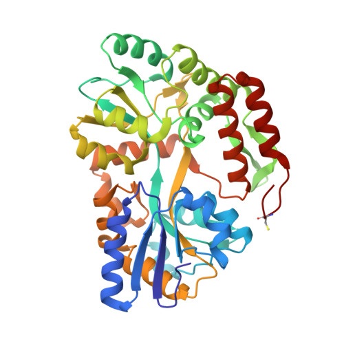 | |
UniProt | |||||
Find proteins for P0AEX9 (Escherichia coli (strain K12)) Explore P0AEX9 Go to UniProtKB: P0AEX9 | |||||
Entity Groups | |||||
| Sequence Clusters | 30% Identity50% Identity70% Identity90% Identity95% Identity100% Identity | ||||
| UniProt Group | P0AEX9 | ||||
Sequence AnnotationsExpand | |||||
| |||||
Find similar proteins by: Sequence | 3D Structure
Entity ID: 2 | |||||
|---|---|---|---|---|---|
| Molecule | Chains | Sequence Length | Organism | Details | Image |
| Ristocetin | 7 | Amycolatopsis lurida | Mutation(s): 0 | 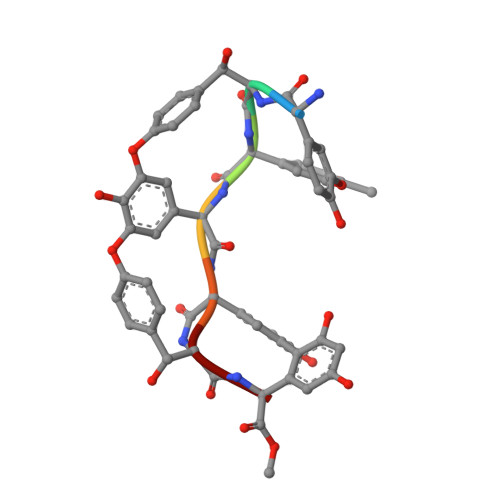 | |
Sequence AnnotationsExpand | |||||
| |||||
Oligosaccharides


Small Molecules
| Ligands 4 Unique | |||||
|---|---|---|---|---|---|
| ID | Chains | Name / Formula / InChI Key | 2D Diagram | 3D Interactions | |
| MAN Query on MAN | N [auth B], P [auth C] | alpha-D-mannopyranose C6 H12 O6 WQZGKKKJIJFFOK-PQMKYFCFSA-N |  | ||
| RST Query on RST | M [auth B], O [auth C] | 3-amino-2,3,6-trideoxy-alpha-L-ribo-hexopyranose C6 H13 N O3 BBMKQGIZNKEDOX-KCDKBNATSA-N |  | ||
| SO4 Query on SO4 | J [auth A], K [auth A], L [auth A], Q [auth C] | SULFATE ION O4 S QAOWNCQODCNURD-UHFFFAOYSA-L |  | ||
| IPA Query on IPA | G [auth A], H [auth A], I [auth A] | ISOPROPYL ALCOHOL C3 H8 O KFZMGEQAYNKOFK-UHFFFAOYSA-N |  | ||
| Modified Residues 3 Unique | |||||
|---|---|---|---|---|---|
| ID | Chains | Type | Formula | 2D Diagram | Parent |
| CCS Query on CCS | A | L-PEPTIDE LINKING | C5 H9 N O4 S |  | CYS |
| MDF Query on MDF | B, C | L-PEPTIDE LINKING | C9 H11 N O4 |  | TYR |
| OMX Query on OMX | B, C | L-PEPTIDE LINKING | C9 H11 N O4 |  | TYR |
Biologically Interesting Molecules (External Reference) 1 Unique
Entity ID: 4 | |||||
|---|---|---|---|---|---|
| ID | Chains | Name | Type/Class | 2D Diagram | 3D Interactions |
| PRD_900001 Query on PRD_900001 | F | alpha-maltose | Oligosaccharide / Nutrient |  | |
Experimental Data & Validation
Experimental Data
- Method: X-RAY DIFFRACTION
- Resolution: 1.85 Å
- R-Value Free: 0.219
- R-Value Work: 0.176
- R-Value Observed: 0.178
- Space Group: P 21 21 21
Unit Cell:
| Length ( Å ) | Angle ( ˚ ) |
|---|---|
| a = 57.833 | α = 90 |
| b = 75.005 | β = 90 |
| c = 90.778 | γ = 90 |
| Software Name | Purpose |
|---|---|
| XSCALE | data scaling |
| MOLREP | phasing |
| PHENIX | refinement |
| PDB_EXTRACT | data extraction |
| XDS | data reduction |
Entry History
Deposition Data
- Released Date: 2012-06-06 Deposition Author(s): Economou, N.J., Weeks, S.D., Grasty, K.C., Nahoum, V., Loll, P.J.
Revision History (Full details and data files)
- Version 1.0: 2012-06-06
Type: Initial release - Version 1.1: 2016-02-17
Changes: Derived calculations, Non-polymer description - Version 2.0: 2020-07-29
Type: Remediation
Reason: Carbohydrate remediation
Changes: Atomic model, Data collection, Database references, Derived calculations, Non-polymer description, Polymer sequence, Structure summary - Version 2.1: 2023-09-13
Changes: Data collection, Database references, Refinement description, Structure summary - Version 2.2: 2023-12-06
Changes: Data collection, Derived calculations
