3RY2

Wild-type core streptavidin-biotin complex at atomic resolution


Experimental Data Snapshot

  • Method: X-RAY DIFFRACTION
  • Resolution: 0.95 Å
  • R-Value Free: 0.131 
  • R-Value Work: 0.115 
  • R-Value Observed: 0.116 

wwPDB Validation   3D Report Full Report


Ligand Structure Quality Assessment 


This is version 1.3 of the entry. See complete history


Literature

Streptavidin and its biotin complex at atomic resolution.

Le Trong, I.Wang, Z.Hyre, D.E.Lybrand, T.P.Stayton, P.S.Stenkamp, R.E.

(2011) Acta Crystallogr D Biol Crystallogr 67: 813-821

  • DOI: https://doi.org/10.1107/S0907444911027806
  • Primary Citation of Related Structures:  
    3RY1, 3RY2

  • PubMed Abstract: 

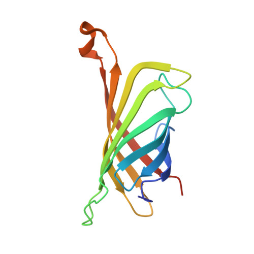
    Atomic resolution crystallographic studies of streptavidin and its biotin complex have been carried out at 1.03 and 0.95 Å, respectively. The wild-type protein crystallized with a tetramer in the asymmetric unit, while the crystals of the biotin complex contained two subunits in the asymmetric unit. Comparison of the six subunits shows the various ways in which the protein accommodates ligand binding and different crystal-packing environments. Conformational variation is found in each of the polypeptide loops connecting the eight strands in the β-sandwich subunit, but the largest differences are found in the flexible binding loop (residues 45-52). In three of the unliganded subunits the loop is in an `open' conformation, while in the two subunits binding biotin, as well as in one of the unliganded subunits, this loop `closes' over the biotin-binding site. The `closed' loop contributes to the protein's high affinity for biotin. Analysis of the anisotropic displacement parameters included in the crystallographic models is consistent with the variation found in the loop structures and the view that the dynamic nature of the protein structure contributes to the ability of the protein to bind biotin so tightly.


  • Organizational Affiliation

    Department of Biological Structure, University of Washington, Seattle, USA.


Macromolecules
Find similar proteins by:  (by identity cutoff)  |  3D Structure
Entity ID: 1
MoleculeChains Sequence LengthOrganismDetailsImage
Streptavidin
A, B
127Streptomyces avidiniiMutation(s): 0 
UniProt
Find proteins for P22629 (Streptomyces avidinii)
Explore P22629 
Go to UniProtKB:  P22629
Entity Groups  
Sequence Clusters30% Identity50% Identity70% Identity90% Identity95% Identity100% Identity
UniProt GroupP22629
Sequence Annotations
Expand
  • Reference Sequence
Experimental Data & Validation

Experimental Data

  • Method: X-RAY DIFFRACTION
  • Resolution: 0.95 Å
  • R-Value Free: 0.131 
  • R-Value Work: 0.115 
  • R-Value Observed: 0.116 
  • Space Group: I 2 2 2
Unit Cell:
Length ( Å )Angle ( ˚ )
a = 46.426α = 90
b = 94.062β = 90
c = 104.608γ = 90
Software Package:
Software NamePurpose
DENZOdata reduction
SCALEPACKdata scaling
REFMACrefinement
PDB_EXTRACTdata extraction
Blu-Icedata collection
Fcphasing

Structure Validation

View Full Validation Report


Ligand Structure Quality Assessment 


Entry History 

Deposition Data

Revision History  (Full details and data files)

  • Version 1.0: 2011-08-24
    Type: Initial release
  • Version 1.1: 2013-06-19
    Changes: Database references
  • Version 1.2: 2017-11-08
    Changes: Refinement description
  • Version 1.3: 2023-09-13
    Changes: Data collection, Database references, Derived calculations, Refinement description