Second-Contact Shell Mutation Diminishes Streptavidin-Biotin Binding Affinity through Transmitted Effects on Equilibrium Dynamics.
Baugh, L., Le Trong, I., Cerutti, D.S., Mehta, N., Gulich, S., Stayton, P.S., Stenkamp, R.E., Lybrand, T.P.(2012) Biochemistry 51: 597-607
- PubMed: 22145986
- DOI: https://doi.org/10.1021/bi201221j
- Primary Citation of Related Structures:
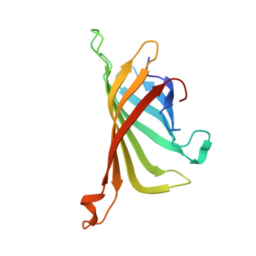
3T6F, 3T6L - PubMed Abstract:
We report a point mutation in the second contact shell of the high-affinity streptavidin-biotin complex that appears to reduce binding affinity through transmitted effects on equilibrium dynamics. The Y54F streptavidin mutation causes a 75-fold loss of binding affinity with 73-fold faster dissociation, a large loss of binding enthalpy (ΔΔH = 3.4 kcal/mol at 37 °C), and a small gain in binding entropy (TΔΔS = 0.7 kcal/mol). The removed Y54 hydroxyl is replaced by a water molecule in the bound structure, but there are no observable changes in structure in the first contact shell and no additional changes surrounding the mutation. Molecular dynamics simulations reveal a large increase in the atomic fluctuation amplitudes for W79, a key biotin contact residue, compared to the fluctuation amplitudes in the wild-type. The increased W79 atomic fluctuation amplitudes are caused by loss of water-mediated hydrogen bonds between the Y54 hydroxyl group and peptide backbone atoms in and near W79. We propose that the increased atomic fluctuation amplitudes diminish the integrity of the W79-biotin interaction and represents a loosening of the "tryptophan collar" that is critical to the slow dissociation and high affinity of streptavidin-biotin binding. These results illustrate how changes in protein dynamics distal to the ligand binding pocket can have a profound impact on ligand binding, even when equilibrium structure is unperturbed.
Organizational Affiliation:
Department of Bioengineering, University of Washington, Seattle, Washington 98195, United States.