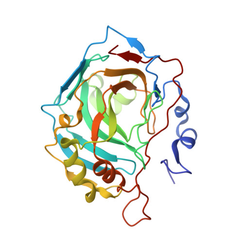
3TMJ

Joint X-ray/neutron structure of human carbonic anhydrase II at pH 7.8


Experimental Data Snapshot

  • Method: NEUTRON DIFFRACTION
  • Resolution: 2.00 Å
  • R-Value Free: 0.294 
  • R-Value Work: 0.280 

  • Method: X-RAY DIFFRACTION

wwPDB Validation   3D Report Full Report


This is version 1.4 of the entry. See complete history


Literature

Neutron Structure of Human Carbonic Anhydrase II: A Hydrogen-Bonded Water Network "Switch" Is Observed between pH 7.8 and 10.0.

Fisher, Z.Kovalevsky, A.Y.Mustyakimov, M.Silverman, D.N.McKenna, R.Langan, P.

(2011) Biochemistry 50: 9421-9423

  • DOI: https://doi.org/10.1021/bi201487b

  • PubMed Abstract: 

    The neutron structure of wild-type human carbonic anhydrase II at pH 7.8 has been determined to 2.0 Å resolution. Detailed analysis and comparison to the previously determined structure at pH 10.0 show important differences in the protonation of key catalytic residues in the active site as well as a rearrangement of the H-bonded water network. For the first time, a completed H-bonded network stretching from the Zn-bound solvent to the proton shuttling residue, His64, has been directly observed.


  • Organizational Affiliation

    Bioscience Division, Los Alamos National Laboratory, Los Alamos, New Mexico 87544, United States. zfisher@lanl.gov


Macromolecules
Find similar proteins by:  (by identity cutoff)  |  3D Structure
Entity ID: 1
MoleculeChains Sequence LengthOrganismDetailsImage
Carbonic anhydrase 2258Homo sapiensMutation(s): 0 
Gene Names: CA2
EC: 4.2.1.1
UniProt & NIH Common Fund Data Resources
Find proteins for P00918 (Homo sapiens)
Explore P00918 
Go to UniProtKB:  P00918
PHAROS:  P00918
GTEx:  ENSG00000104267 
Entity Groups  
Sequence Clusters30% Identity50% Identity70% Identity90% Identity95% Identity100% Identity
UniProt GroupP00918
Sequence Annotations
Expand
  • Reference Sequence
Small Molecules
Ligands 1 Unique
IDChains Name / Formula / InChI Key2D Diagram3D Interactions
ZN
Query on ZN

Download Ideal Coordinates CCD File 
B [auth A]ZINC ION
Zn
PTFCDOFLOPIGGS-UHFFFAOYSA-N
Experimental Data & Validation

Experimental Data

  • Method: NEUTRON DIFFRACTION
  • Resolution: 2.00 Å
  • R-Value Free: 0.294 
  • R-Value Work: 0.280 
  • Space Group: P 1 21 1
Unit Cell:
Length ( Å )Angle ( ˚ )
a = 42.83α = 90
b = 41.7β = 104.58
c = 72.92γ = 90
Software Package:
Software NamePurpose
PCSdata collection
nCNSrefinement
d*TREKdata reduction
SCALAdata scaling
d*TREKdata scaling
nCNSphasing

Structure Validation

View Full Validation Report


Entry History 

Deposition Data

  • Released Date: 2011-11-09 
  • Deposition Author(s): Fisher, Z.

Revision History  (Full details and data files)

  • Version 1.0: 2011-11-09
    Type: Initial release
  • Version 1.1: 2017-11-08
    Changes: Refinement description
  • Version 1.2: 2018-04-25
    Changes: Data collection
  • Version 1.3: 2018-06-13
    Changes: Data collection
  • Version 1.4: 2023-09-13
    Changes: Data collection, Database references, Derived calculations, Refinement description