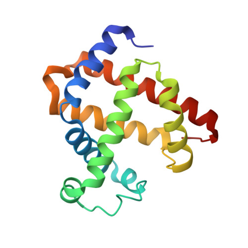
Complex of myoglobin with phenol bound in a proximal cavity.
Huang, X., Wang, C., Celeste, L.R., Lovelace, L.L., Sun, S., Dawson, J.H., Lebioda, L.(2012) Acta Crystallogr Sect F Struct Biol Cryst Commun 68: 1465-1471
- PubMed: 23192025
- DOI: https://doi.org/10.1107/S1744309112045514
- Primary Citation of Related Structures:
3U3E, 4H07, 4H0B - PubMed Abstract:
Sperm whale myoglobin (Mb) has weak dehaloperoxidase activity and catalyzes the peroxidative dehalogenation of 2,4,6-trichlorophenol (TCP) to 2,6-dichloroquinone. Crystals of Mb and of its more active G65T variant were used to study the binding of TCP, 4-iodophenol (4-IP) and phenol. The structures of crystals soaked overnight in a 10 mM solution of phenol revealed that a phenol molecule binds in the proximal cavity, forming a hydrogen bond to the hydroxyl of Tyr146 and hydrophobic contacts which include interactions with Cβ and Cγ of the proximal histidine His93. The phenol position corresponds to the strongest xenon binding site, Xe1. It appears that the ligand enters the proximal cavity through a gate formed by the flexible loops 79-86 and 93-103. TCP and 4-IP do not bind to Mb in this manner under similar conditions; however, it appears to be likely that dimethyl sulfoxide (DMSO), which was used at a concentration of 0.8 M to facilitate 4-IP dissolution, binds in the phenol/Xe1 binding site. In this structure, a water molecule coordinated to the heme iron was replaced by an oxygen molecule, reflecting the reduction of the heme. Crystals of Mb and G65T Mb soaked for 5-10 min did not show bound phenol. Kinetic studies of TCP dechlorination showed that phenol has a dual effect: it acts as a competitive inhibitor that is likely to interfere with TCP binding at the heme edge and as a weak activator, likely through binding in the proximal cavity. The lack of phenol bound at the heme edge in the crystal structures suggests that its inhibitory binding only takes place when the heme is activated by hydrogen peroxide.
Organizational Affiliation:
Department of Chemistry and Biochemistry, University of South Carolina, Columbia, SC 20208, USA.