3V3D

Crystal Structure of an eYFP single mutant


Experimental Data Snapshot

  • Method: X-RAY DIFFRACTION
  • Resolution: 1.95 Å
  • R-Value Free: 0.229 
  • R-Value Work: 0.184 
  • R-Value Observed: 0.187 

wwPDB Validation   3D Report Full Report


This is version 1.5 of the entry. See complete history


Literature

Improving the second-order nonlinear optical response of fluorescent proteins: the symmetry argument.

De Meulenaere, E.Nguyen Bich, N.de Wergifosse, M.Van Hecke, K.Van Meervelt, L.Vanderleyden, J.Champagne, B.Clays, K.

(2013) J Am Chem Soc 135: 4061-4069

  • DOI: https://doi.org/10.1021/ja400098b
  • Primary Citation of Related Structures:  
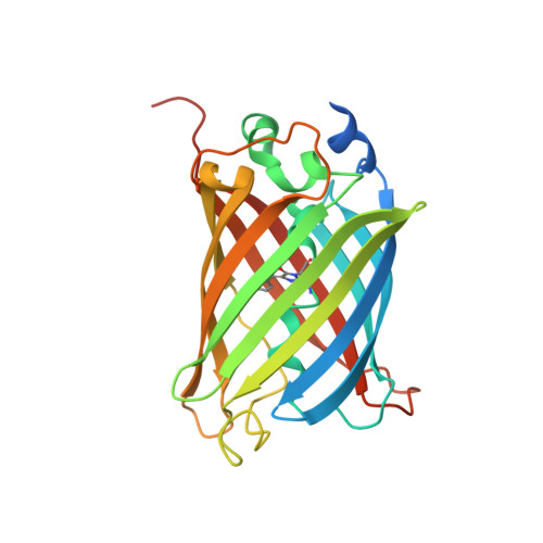
    3V3D

  • PubMed Abstract: 

    We have successfully designed and expressed a new fluorescent protein with improved second-order nonlinear optical properties. It is the first time that a fluorescent protein has been rationally altered for this particular characteristic. On the basis of the specific noncentrosymmetry requirements for second-order nonlinear optical effects, we had hypothesized that the surprisingly low first hyperpolarizability (β) of the enhanced yellow fluorescent protein (eYFP) could be explained by centrosymmetric stacking of the chromophoric Tyr66 and the neighboring Tyr203 residue. The inversion center was removed by mutating Tyr203 into Phe203, with minor changes in the linear optical properties and even an improved fluorescence quantum yield. Structure determination by X-ray crystallography as well as linear optical characterization corroborate a correct folding and maturation. Measurement of β by means of hyper-Rayleigh scattering (HRS) as well as their analysis using quantum chemistry calculations validate our hypothesis. This observation can eventually lead to improved red fluorescent proteins for even better performance. On the basis of the specific function (second-harmonic generation), the color of its emission, and in analogy with the "fruit" names, we propose SHardonnay as the name for this Tyr203Phe mutant of eYFP.


  • Organizational Affiliation

    Centre of Microbial and Plant Genetics, KU Leuven, Kasteelpark Arenberg 20, BE-3001 Leuven, Belgium.


Macromolecules
Find similar proteins by:  (by identity cutoff)  |  3D Structure
Entity ID: 1
MoleculeChains Sequence LengthOrganismDetailsImage
Yellow fluorescent protein257Aequorea victoriaMutation(s): 5 
Gene Names: GFP
UniProt
Find proteins for P42212 (Aequorea victoria)
Explore P42212 
Go to UniProtKB:  P42212
Entity Groups  
Sequence Clusters30% Identity50% Identity70% Identity90% Identity95% Identity100% Identity
UniProt GroupP42212
Sequence Annotations
Expand
  • Reference Sequence
Small Molecules
Ligands 1 Unique
IDChains Name / Formula / InChI Key2D Diagram3D Interactions
SO4
Query on SO4

Download Ideal Coordinates CCD File 
B [auth A]SULFATE ION
O4 S
QAOWNCQODCNURD-UHFFFAOYSA-L
Modified Residues  1 Unique
IDChains TypeFormula2D DiagramParent
CR2
Query on CR2
A
L-PEPTIDE LINKINGC13 H13 N3 O4GLY, TYR, GLY
Experimental Data & Validation

Experimental Data

  • Method: X-RAY DIFFRACTION
  • Resolution: 1.95 Å
  • R-Value Free: 0.229 
  • R-Value Work: 0.184 
  • R-Value Observed: 0.187 
  • Space Group: P 41 21 2
Unit Cell:
Length ( Å )Angle ( ˚ )
a = 59.858α = 90
b = 59.858β = 90
c = 165.54γ = 90
Software Package:
Software NamePurpose
PHASERphasing
PHENIXrefinement
XDSdata reduction
SCALAdata scaling

Structure Validation

View Full Validation Report


Entry History 

Deposition Data

Revision History  (Full details and data files)

  • Version 1.0: 2012-12-26
    Type: Initial release
  • Version 1.1: 2013-03-06
    Changes: Database references
  • Version 1.2: 2013-03-27
    Changes: Database references
  • Version 1.3: 2017-11-08
    Changes: Refinement description
  • Version 1.4: 2023-09-13
    Changes: Data collection, Database references, Derived calculations, Refinement description
  • Version 1.5: 2023-12-06
    Changes: Data collection