3W15

Structure of peroxisomal targeting signal 2 (PTS2) of Saccharomyces cerevisiae 3-ketoacyl-CoA thiolase in complex with Pex7p and Pex21p


Experimental Data Snapshot

  • Method: X-RAY DIFFRACTION
  • Resolution: 1.80 Å
  • R-Value Free: 0.224 
  • R-Value Work: 0.189 
  • R-Value Observed: 0.191 

wwPDB Validation   3D Report Full Report


This is version 1.2 of the entry. See complete history


Literature

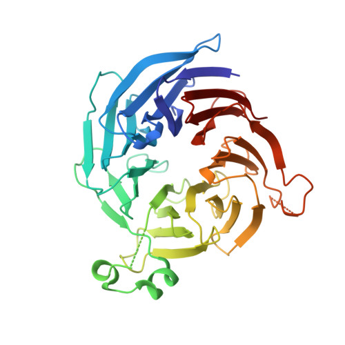
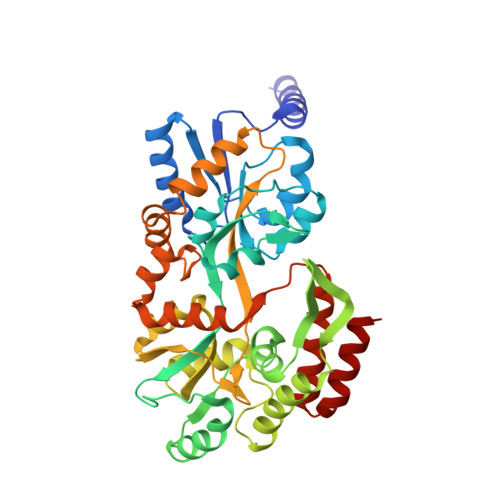
Crystal structure of peroxisomal targeting signal-2 bound to its receptor complex Pex7p-Pex21p

Pan, D.Nakatsu, T.Kato, H.

(2013) Nat Struct Mol Biol 20: 987-993

  • DOI: https://doi.org/10.1038/nsmb.2618
  • Primary Citation of Related Structures:  
    3W15

  • PubMed Abstract: 

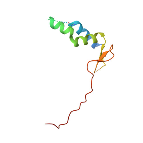
    Appropriate targeting of matrix proteins to peroxisomes is mainly directed by two types of peroxisomal targeting signals, PTS1 and PTS2. Although the basis of PTS1 recognition has been revealed by structural studies, that of PTS2 recognition remains elusive. Here we present the crystal structure of a heterotrimeric PTS2-recognition complex from Saccharomyces cerevisiae, containing Pex7p, the C-terminal region of Pex21p and the PTS2 of the peroxisomal 3-ketoacyl-CoA thiolase. Pex7p forms a β-propeller structure and provides a platform for cooperative interactions with both the amphipathic PTS2 helix and Pex21p. The C-terminal region of Pex21p directly covers the hydrophobic surfaces of both Pex7p and PTS2, and the resulting hydrophobic core is the primary determinant of stable complex formation. Together with in vivo and in vitro functional assays of Pex7p and Pex21p variants, our findings reveal the molecular mechanism of PTS2 recognition.


  • Organizational Affiliation

    Department of Structural Biology, Graduate School of Pharmaceutical Sciences, Kyoto University, Kyoto, Japan. pan.dongqing@pharm.kyoto-u.ac.jp


Macromolecules
Find similar proteins by:  (by identity cutoff)  |  3D Structure
Entity ID: 1
MoleculeChains Sequence LengthOrganismDetailsImage
Peroxisomal targeting signal 2 receptor368Saccharomyces cerevisiae S288CMutation(s): 0 
Gene Names: PEX7
UniProt
Find proteins for P39108 (Saccharomyces cerevisiae (strain ATCC 204508 / S288c))
Explore P39108 
Go to UniProtKB:  P39108
Entity Groups  
Sequence Clusters30% Identity50% Identity70% Identity90% Identity95% Identity100% Identity
UniProt GroupP39108
Sequence Annotations
Expand
  • Reference Sequence
Find similar proteins by:  (by identity cutoff)  |  3D Structure
Entity ID: 2
MoleculeChains Sequence LengthOrganismDetailsImage
Peroxisomal membrane protein PEX21101Saccharomyces cerevisiae S288CMutation(s): 0 
Gene Names: PEX21
UniProt
Find proteins for P50091 (Saccharomyces cerevisiae (strain ATCC 204508 / S288c))
Explore P50091 
Go to UniProtKB:  P50091
Entity Groups  
Sequence Clusters30% Identity50% Identity70% Identity90% Identity95% Identity100% Identity
UniProt GroupP50091
Sequence Annotations
Expand
  • Reference Sequence
Find similar proteins by:  (by identity cutoff)  |  3D Structure
Entity ID: 3
MoleculeChains Sequence LengthOrganismDetailsImage
3-ketoacyl-CoA thiolase, peroxisomal, Maltose-binding periplasmic protein389Saccharomyces cerevisiae S288CEscherichia coli K-12
This entity is chimeric
Mutation(s): 0 
Gene Names: FOX3
UniProt
Find proteins for P27796 (Saccharomyces cerevisiae (strain ATCC 204508 / S288c))
Explore P27796 
Go to UniProtKB:  P27796
Find proteins for P0AEX9 (Escherichia coli (strain K12))
Explore P0AEX9 
Go to UniProtKB:  P0AEX9
Entity Groups  
Sequence Clusters30% Identity50% Identity70% Identity90% Identity95% Identity100% Identity
UniProt GroupsP0AEX9P27796
Sequence Annotations
Expand
  • Reference Sequence
Small Molecules
Ligands 2 Unique
IDChains Name / Formula / InChI Key2D Diagram3D Interactions
NO3
Query on NO3

Download Ideal Coordinates CCD File 
D [auth A]
E [auth A]
G [auth B]
H [auth C]
I [auth C]
D [auth A],
E [auth A],
G [auth B],
H [auth C],
I [auth C],
J [auth C],
K [auth C],
L [auth C],
M [auth C]
NITRATE ION
N O3
NHNBFGGVMKEFGY-UHFFFAOYSA-N
MG
Query on MG

Download Ideal Coordinates CCD File 
F [auth A]
N [auth C]
O [auth C]
P [auth C]
Q [auth C]
F [auth A],
N [auth C],
O [auth C],
P [auth C],
Q [auth C],
R [auth C]
MAGNESIUM ION
Mg
JLVVSXFLKOJNIY-UHFFFAOYSA-N
Experimental Data & Validation

Experimental Data

  • Method: X-RAY DIFFRACTION
  • Resolution: 1.80 Å
  • R-Value Free: 0.224 
  • R-Value Work: 0.189 
  • R-Value Observed: 0.191 
  • Space Group: P 1 21 1
Unit Cell:
Length ( Å )Angle ( ˚ )
a = 78.89α = 90
b = 52.75β = 106.59
c = 97.57γ = 90
Software Package:
Software NamePurpose
MOSFLMdata reduction
SCALAdata scaling
PHASERphasing
REFMACrefinement
PDB_EXTRACTdata extraction

Structure Validation

View Full Validation Report


Entry History 

Deposition Data

Revision History  (Full details and data files)

  • Version 1.0: 2013-07-03
    Type: Initial release
  • Version 1.1: 2013-10-16
    Changes: Database references
  • Version 1.2: 2017-08-16
    Changes: Refinement description, Source and taxonomy