3X1U

Crystal structure of nucleosome core particle in the presence of histone variants involved in reprogramming


Experimental Data Snapshot

  • Method: X-RAY DIFFRACTION
  • Resolution: 3.25 Å
  • R-Value Free: 0.270 
  • R-Value Work: 0.200 
  • R-Value Observed: 0.204 

wwPDB Validation   3D Report Full Report


This is version 1.2 of the entry. See complete history


Literature

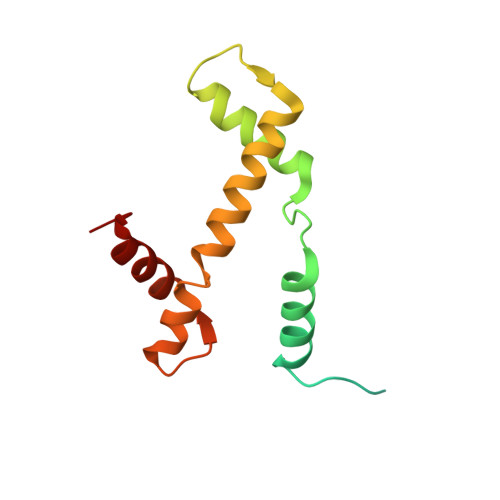
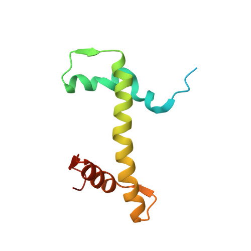
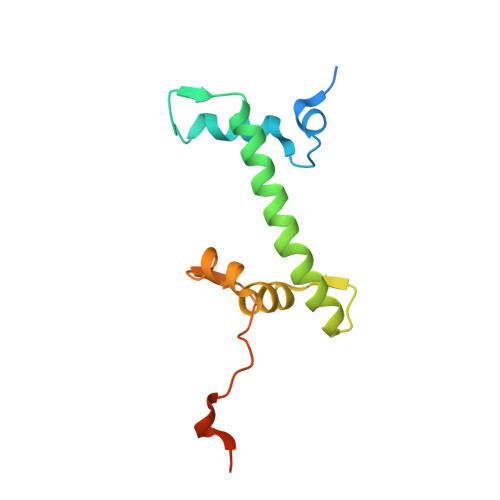
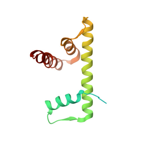
Structural and functional analyses of nucleosome complexes with mouse histone variants TH2a and TH2b, involved in reprogramming.

Padavattan, S.Shinagawa, T.Hasegawa, K.Kumasaka, T.Ishii, S.Kumarevel, T.

(2015) Biochem Biophys Res Commun 464: 929-935

  • DOI: https://doi.org/10.1016/j.bbrc.2015.07.070
  • Primary Citation of Related Structures:  
    3X1S, 3X1T, 3X1U, 3X1V

  • PubMed Abstract: 

    Histone variants TH2a and TH2b are highly expressed in testes, oocytes and zygotes. Our recent analysis suggested that these histone variants enhance the induced generation of pluripotent stem cells (iPSCs) when co-expressed along with four transcription factors, Oct3/4, Sox2, Klf4 and c-Myc (OSKM), and are associated with an open chromatin structure [1]. In the present study, we report the crystal structures of nucleosomes (NCPs) with the mouse histone variants, TH2a and TH2b. The structures revealed two significant changes, as compared to the canonical counterparts: fewer histone-DNA contacts and changes in dimer-dimer interactions between TH2a-TH2a' (L1-loop). In vivo studies with domain swapping and point mutants of the variants revealed that the residues in the histone tails and the TH2a-L1 loop are important for reprogramming. Taken together, our work indicates that the NCP variants with structural modifications and flexible tails are most likely important for enhanced reprogramming of functions.


  • Organizational Affiliation

    RIKEN SPring-8 Center, Harima Institute, 1-1-1 Kouto, Sayo, Hyogo 679-5148, Japan.


Macromolecules

Find similar proteins by:  (by identity cutoff)  |  3D Structure
Entity ID: 2
MoleculeChains Sequence LengthOrganismDetailsImage
Histone H3.1C [auth A],
G [auth E]
135Homo sapiensMutation(s): 0 
Gene Names: H3.1
UniProt & NIH Common Fund Data Resources
Find proteins for P68431 (Homo sapiens)
Explore P68431 
Go to UniProtKB:  P68431
PHAROS:  P68431
Entity Groups  
Sequence Clusters30% Identity50% Identity70% Identity90% Identity95% Identity100% Identity
UniProt GroupP68431
Sequence Annotations
Expand
  • Reference Sequence
Find similar proteins by:  (by identity cutoff)  |  3D Structure
Entity ID: 3
MoleculeChains Sequence LengthOrganismDetailsImage
Histone H4D [auth B],
H [auth F]
102Homo sapiensMutation(s): 0 
Gene Names: H4
UniProt & NIH Common Fund Data Resources
Find proteins for P62805 (Homo sapiens)
Explore P62805 
Go to UniProtKB:  P62805
PHAROS:  P62805
Entity Groups  
Sequence Clusters30% Identity50% Identity70% Identity90% Identity95% Identity100% Identity
UniProt GroupP62805
Sequence Annotations
Expand
  • Reference Sequence
Find similar proteins by:  (by identity cutoff)  |  3D Structure
Entity ID: 4
MoleculeChains Sequence LengthOrganismDetailsImage
Histone H2AE [auth C],
I [auth G]
128Mus musculusMutation(s): 0 
Gene Names: H2AA
UniProt
Find proteins for Q8CGP4 (Mus musculus)
Explore Q8CGP4 
Go to UniProtKB:  Q8CGP4
Entity Groups  
Sequence Clusters30% Identity50% Identity70% Identity90% Identity95% Identity100% Identity
UniProt GroupQ8CGP4
Sequence Annotations
Expand
  • Reference Sequence
Find similar proteins by:  (by identity cutoff)  |  3D Structure
Entity ID: 5
MoleculeChains Sequence LengthOrganismDetailsImage
Histone H2B type 1-BF [auth D],
J [auth H]
124Homo sapiensMutation(s): 0 
Gene Names: H2B
UniProt & NIH Common Fund Data Resources
Find proteins for P33778 (Homo sapiens)
Explore P33778 
Go to UniProtKB:  P33778
PHAROS:  P33778
GTEx:  ENSG00000276410 
Entity Groups  
Sequence Clusters30% Identity50% Identity70% Identity90% Identity95% Identity100% Identity
UniProt GroupP33778
Sequence Annotations
Expand
  • Reference Sequence
Find similar nucleic acids by:  (by identity cutoff)  |  3D Structure
Entity ID: 1
MoleculeChains LengthOrganismImage
DNA (146-MER)A [auth I],
B [auth J]
146synthetic construct
Sequence Annotations
Expand
  • Reference Sequence
Small Molecules
Ligands 2 Unique
IDChains Name / Formula / InChI Key2D Diagram3D Interactions
MN
Query on MN

Download Ideal Coordinates CCD File 
K [auth I],
L [auth I],
R [auth E],
T [auth G]
MANGANESE (II) ION
Mn
WAEMQWOKJMHJLA-UHFFFAOYSA-N
CL
Query on CL

Download Ideal Coordinates CCD File 
M [auth I]
N [auth I]
O [auth I]
P [auth J]
Q [auth J]
M [auth I],
N [auth I],
O [auth I],
P [auth J],
Q [auth J],
S [auth E]
CHLORIDE ION
Cl
VEXZGXHMUGYJMC-UHFFFAOYSA-M
Experimental Data & Validation

Experimental Data

  • Method: X-RAY DIFFRACTION
  • Resolution: 3.25 Å
  • R-Value Free: 0.270 
  • R-Value Work: 0.200 
  • R-Value Observed: 0.204 
  • Space Group: P 21 21 21
Unit Cell:
Length ( Å )Angle ( ˚ )
a = 104.427α = 90
b = 109.184β = 90
c = 175.001γ = 90
Software Package:
Software NamePurpose
PHENIXrefinement
PDB_EXTRACTdata extraction
HKL-2000data collection
HKL-2000data reduction
HKL-2000data scaling
PHASERphasing
MOLREPphasing

Structure Validation

View Full Validation Report


Entry History 

Deposition Data

Revision History  (Full details and data files)

  • Version 1.0: 2015-09-23
    Type: Initial release
  • Version 1.1: 2017-11-22
    Changes: Refinement description
  • Version 1.2: 2023-11-08
    Changes: Data collection, Database references, Derived calculations, Refinement description