4AR7

X-ray structure of the cyan fluorescent protein mTurquoise


Experimental Data Snapshot

  • Method: X-RAY DIFFRACTION
  • Resolution: 1.23 Å
  • R-Value Free: 0.166 
  • R-Value Work: 0.143 
  • R-Value Observed: 0.144 

wwPDB Validation   3D Report Full Report


This is version 1.4 of the entry. See complete history


Literature

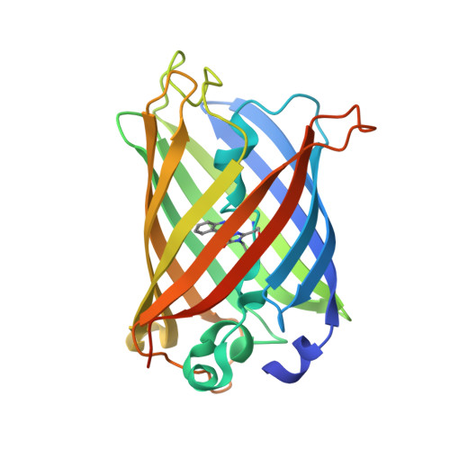
Structure of a Fluorescent Protein from Aequorea Victoria Bearing the Obligate-Monomer Mutation A206K.

von Stetten, D.Noirclerc-Savoye, M.Goedhart, J.Gadella, T.W.J.J.Royant, A.

(2012) Acta Crystallogr Sect F Struct Biol Cryst Commun 68: 878

  • DOI: https://doi.org/10.1107/S1744309112028667
  • Primary Citation of Related Structures:  
    4AR7

  • PubMed Abstract: 

    The green fluorescent protein (GFP) from the jellyfish Aequoria victoria has been shown to dimerize at high concentrations, which could lead to artefacts in imaging experiments. To ensure a truly monomeric state, an A206K mutation has been introduced into most of its widely used variants, with minimal effect on the spectroscopic properties. Here, the first structure of one of these variants, the cyan fluorescent protein mTurquoise, is presented and compared with that of its dimeric version mTurquoise-K206A. No significant structural change is detected in the chromophore cavity, reinforcing the notion that this mutation is spectroscopically silent and validating that the structural analysis performed on dimeric mutants also applies to monomeric versions. Finally, it is explained why cyan versions of GFP containing the Y66W and N146I mutations do not require the A206K mutation to prevent dimerization at high concentrations.


  • Organizational Affiliation

    Structural Biology Group, European Synchrotron Radiation Facility, 6 Rue Jules Horowitz, 38043 Grenoble, France.


Macromolecules
Find similar proteins by:  (by identity cutoff)  |  3D Structure
Entity ID: 1
MoleculeChains Sequence LengthOrganismDetailsImage
GREEN FLUORESCENT PROTEIN243Aequorea victoriaMutation(s): 10 
UniProt
Find proteins for P42212 (Aequorea victoria)
Explore P42212 
Go to UniProtKB:  P42212
Entity Groups  
Sequence Clusters30% Identity50% Identity70% Identity90% Identity95% Identity100% Identity
UniProt GroupP42212
Sequence Annotations
Expand
  • Reference Sequence
Small Molecules
Modified Residues  1 Unique
IDChains TypeFormula2D DiagramParent
SWG
Query on SWG
A
L-PEPTIDE LINKINGC16 H16 N4 O4SER, TRP, GLY
Experimental Data & Validation

Experimental Data

  • Method: X-RAY DIFFRACTION
  • Resolution: 1.23 Å
  • R-Value Free: 0.166 
  • R-Value Work: 0.143 
  • R-Value Observed: 0.144 
  • Space Group: P 21 21 21
Unit Cell:
Length ( Å )Angle ( ˚ )
a = 51.452α = 90
b = 62.558β = 90
c = 69.543γ = 90
Software Package:
Software NamePurpose
REFMACrefinement
XDSdata reduction
SCALAdata scaling
REFMACphasing

Structure Validation

View Full Validation Report


Entry History 

Deposition Data

Revision History  (Full details and data files)

  • Version 1.0: 2012-08-08
    Type: Initial release
  • Version 1.1: 2012-08-15
    Changes: Database references
  • Version 1.2: 2017-12-20
    Changes: Database references
  • Version 1.3: 2019-10-23
    Changes: Data collection, Database references, Derived calculations, Other
  • Version 1.4: 2023-12-20
    Changes: Data collection, Database references, Derived calculations, Refinement description