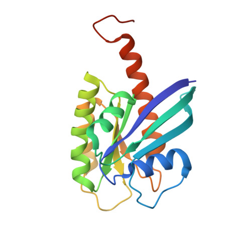
Small-molecule ligands bind to a distinct pocket in Ras and inhibit SOS-mediated nucleotide exchange activity.
Maurer, T., Garrenton, L.S., Oh, A., Pitts, K., Anderson, D.J., Skelton, N.J., Fauber, B.P., Pan, B., Malek, S., Stokoe, D., Ludlam, M.J., Bowman, K.K., Wu, J., Giannetti, A.M., Starovasnik, M.A., Mellman, I., Jackson, P.K., Rudolph, J., Wang, W., Fang, G.(2012) Proc Natl Acad Sci U S A 109: 5299-5304
- PubMed: 22431598
- DOI: https://doi.org/10.1073/pnas.1116510109
- Primary Citation of Related Structures:
4DSN, 4DSO, 4DST, 4DSU - PubMed Abstract:
The Ras gene is frequently mutated in cancer, and mutant Ras drives tumorigenesis. Although Ras is a central oncogene, small molecules that bind to Ras in a well-defined manner and exert inhibitory effects have not been uncovered to date. Through an NMR-based fragment screen, we identified a group of small molecules that all bind to a common site on Ras. High-resolution cocrystal structures delineated a unique ligand-binding pocket on the Ras protein that is adjacent to the switch I/II regions and can be expanded upon compound binding. Structure analysis predicts that compound-binding interferes with the Ras/SOS interactions. Indeed, selected compounds inhibit SOS-mediated nucleotide exchange and prevent Ras activation by blocking the formation of intermediates of the exchange reaction. The discovery of a small-molecule binding pocket on Ras with functional significance provides a new direction in the search of therapeutically effective inhibitors of the Ras oncoprotein.
Organizational Affiliation:
Structural Biology, Genentech, Inc., One DNA Way, South San Francisco, CA 94080, USA.