4G80

Crystal structure of voltage sensing domain of Ci-VSP with fragment antibody (WT, 3.8 A)


Experimental Data Snapshot

  • Method: X-RAY DIFFRACTION
  • Resolution: 3.58 Å
  • R-Value Free: 0.292 
  • R-Value Work: 0.248 
  • R-Value Observed: 0.250 

wwPDB Validation   3D Report Full Report


This is version 1.2 of the entry. See complete history


Literature

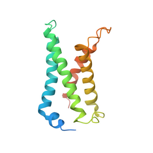
Structural mechanism of voltage-dependent gating in an isolated voltage-sensing domain.

Li, Q.Wanderling, S.Paduch, M.Medovoy, D.Singharoy, A.McGreevy, R.Villalba-Galea, C.A.Hulse, R.E.Roux, B.Schulten, K.Kossiakoff, A.Perozo, E.

(2014) Nat Struct Mol Biol 21: 244-252

  • DOI: https://doi.org/10.1038/nsmb.2768
  • Primary Citation of Related Structures:  
    4G7V, 4G7Y, 4G80

  • PubMed Abstract: 

    The transduction of transmembrane electric fields into protein motion has an essential role in the generation and propagation of cellular signals. Voltage-sensing domains (VSDs) carry out these functions through reorientations of positive charges in the S4 helix. Here, we determined crystal structures of the Ciona intestinalis VSD (Ci-VSD) in putatively active and resting conformations. S4 undergoes an ~5-Å displacement along its main axis, accompanied by an ~60° rotation. This movement is stabilized by an exchange in countercharge partners in helices S1 and S3 that generates an estimated net charge transfer of ~1 eo. Gating charges move relative to a ''hydrophobic gasket' that electrically divides intra- and extracellular compartments. EPR spectroscopy confirms the limited nature of S4 movement in a membrane environment. These results provide an explicit mechanism for voltage sensing and set the basis for electromechanical coupling in voltage-dependent enzymes and ion channels.


  • Organizational Affiliation

    Department of Biochemistry and Molecular Biology, University of Chicago, Chicago, Illinois, USA.


Macromolecules
Find similar proteins by:  (by identity cutoff)  |  3D Structure
Entity ID: 1
MoleculeChains Sequence LengthOrganismDetailsImage
Voltage-sensor containing phosphataseA [auth S],
F [auth T],
K [auth I],
L [auth J]
155Ciona intestinalisMutation(s): 0 
Gene Names: Ci-VSP
Membrane Entity: Yes 
UniProt
Find proteins for F6XHE4 (Ciona intestinalis)
Explore F6XHE4 
Go to UniProtKB:  F6XHE4
Entity Groups  
Sequence Clusters30% Identity50% Identity70% Identity90% Identity95% Identity100% Identity
UniProt GroupF6XHE4
Sequence Annotations
Expand
  • Reference Sequence
Find similar proteins by:  (by identity cutoff)  |  3D Structure
Entity ID: 2
MoleculeChains Sequence LengthOrganismDetailsImage
fragment antibody heavy chainB [auth A],
D [auth C],
G [auth E],
I [auth G]
224Homo sapiensMutation(s): 0 
Entity Groups  
Sequence Clusters30% Identity50% Identity70% Identity90% Identity95% Identity100% Identity
Sequence Annotations
Expand
  • Reference Sequence
Find similar proteins by:  (by identity cutoff)  |  3D Structure
Entity ID: 3
MoleculeChains Sequence LengthOrganismDetailsImage
fragment antibody light chainC [auth B],
E [auth D],
H [auth F],
J [auth H]
211Homo sapiensMutation(s): 0 
Entity Groups  
Sequence Clusters30% Identity50% Identity70% Identity90% Identity95% Identity100% Identity
Sequence Annotations
Expand
  • Reference Sequence
Experimental Data & Validation

Experimental Data

  • Method: X-RAY DIFFRACTION
  • Resolution: 3.58 Å
  • R-Value Free: 0.292 
  • R-Value Work: 0.248 
  • R-Value Observed: 0.250 
  • Space Group: P 1
Unit Cell:
Length ( Å )Angle ( ˚ )
a = 77.29α = 102.63
b = 94.24β = 93.45
c = 193.95γ = 105.25
Software Package:
Software NamePurpose
JBlucedata collection
PHASERphasing
REFMACrefinement
HKL-2000data reduction
HKL-2000data scaling

Structure Validation

View Full Validation Report


Entry History 

Deposition Data

  • Released Date: 2014-02-05 
  • Deposition Author(s): Li, Q.

Revision History  (Full details and data files)

  • Version 1.0: 2014-02-05
    Type: Initial release
  • Version 1.1: 2017-11-15
    Changes: Refinement description
  • Version 1.2: 2017-12-13
    Changes: Database references