4H1P

Use of Europium for SAD Phasing at the Cu K alpha wavelength


Experimental Data Snapshot

  • Method: X-RAY DIFFRACTION
  • Resolution: 2.30 Å
  • R-Value Free: 0.231 
  • R-Value Work: 0.174 
  • R-Value Observed: 0.179 

wwPDB Validation   3D Report Full Report


This is version 1.3 of the entry. See complete history


Literature

Use of europium ions for SAD phasing of Lysozyme at the Cu Kalpha wavelength

Vijayakumar, B.Velmurugan, D.

(2013) Acta Crystallogr Sect F Struct Biol Cryst Commun 69: 20-24

  • DOI: https://doi.org/10.1107/S1744309112047562
  • Primary Citation of Related Structures:  
    4H1P

  • PubMed Abstract: 

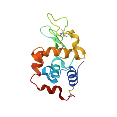
    Europium is shown to be a good anomalous scatterer in SAD phasing for solving the structure of biological macromolecules. The large value of the anomalous contribution of europium, f'' = 11.17 e(-), at the Cu Kα wavelength is an advantage in de novo phasing and automated model building. Tetragonal crystals of hen egg-white lysozyme (HEWL) incorporating europium(III) chloride (50 mM) were obtained which diffracted to a resolution of 2.3 Å at a wavelength of 1.54 Å (Cu Kα). The master data set (360° frames) was split and analyzed for anomalous signal-to-noise ratio, multiplicity, completeness, SAD phasing and automated building. The structure solution and model building of the split data sets were carried out using phenix.autosol and phenix.autobuild. The contributions of the Eu ions to SAD phasing using in-house data collection are discussed. This study revealed successful lysozyme phasing by SAD using laboratory-source data involving Eu ions, which are mainly coordinated by the side chains of Asn46, Asp52 and Asp101 together with some water molecules.


  • Organizational Affiliation

    Centre of Advanced Study in Crystallography and Biophysics, University of Madras, Maraimalai Guindy Campus, Chennai 600 025, India.


Macromolecules
Find similar proteins by:  (by identity cutoff)  |  3D Structure
Entity ID: 1
MoleculeChains Sequence LengthOrganismDetailsImage
Lysozyme C129Gallus gallusMutation(s): 0 
EC: 3.2.1.17
UniProt
Find proteins for P00698 (Gallus gallus)
Explore P00698 
Go to UniProtKB:  P00698
Entity Groups  
Sequence Clusters30% Identity50% Identity70% Identity90% Identity95% Identity100% Identity
UniProt GroupP00698
Sequence Annotations
Expand
  • Reference Sequence
Small Molecules
Ligands 3 Unique
IDChains Name / Formula / InChI Key2D Diagram3D Interactions
EU
Query on EU

Download Ideal Coordinates CCD File 
B [auth A],
C [auth A]
EUROPIUM ION
Eu
MGVUQZZTJGLWJV-UHFFFAOYSA-N
CL
Query on CL

Download Ideal Coordinates CCD File 
D [auth A]
E [auth A]
F [auth A]
G [auth A]
H [auth A]
D [auth A],
E [auth A],
F [auth A],
G [auth A],
H [auth A],
I [auth A],
J [auth A],
K [auth A],
L [auth A],
M [auth A],
N [auth A],
O [auth A],
P [auth A],
Q [auth A]
CHLORIDE ION
Cl
VEXZGXHMUGYJMC-UHFFFAOYSA-M
NA
Query on NA

Download Ideal Coordinates CCD File 
R [auth A]SODIUM ION
Na
FKNQFGJONOIPTF-UHFFFAOYSA-N
Experimental Data & Validation

Experimental Data

  • Method: X-RAY DIFFRACTION
  • Resolution: 2.30 Å
  • R-Value Free: 0.231 
  • R-Value Work: 0.174 
  • R-Value Observed: 0.179 
  • Space Group: P 43 21 2
Unit Cell:
Length ( Å )Angle ( ˚ )
a = 77.485α = 90
b = 77.485β = 90
c = 36.986γ = 90
Software Package:
Software NamePurpose
MAR345dtbdata collection
AutoSolphasing
PHENIXrefinement
AUTOMARdata reduction

Structure Validation

View Full Validation Report


Entry History 

Deposition Data

Revision History  (Full details and data files)

  • Version 1.0: 2012-10-24
    Type: Initial release
  • Version 1.1: 2013-01-09
    Changes: Database references
  • Version 1.2: 2013-04-24
    Changes: Database references
  • Version 1.3: 2020-01-15
    Changes: Derived calculations