


Structural insights of the fluorescent states of CyPet
Liu, R., Zhou, Y.-B., Ding, Y., Hu, X.-J.To be published.
Experimental Data Snapshot
wwPDB Validation 3D Report Full Report

Entity ID: 1 | |||||
|---|---|---|---|---|---|
| Molecule | Chains | Sequence Length | Organism | Details | Image |
| Green fluorescent protein | 237 | Aequorea victoria | Mutation(s): 8 Gene Names: GFP | 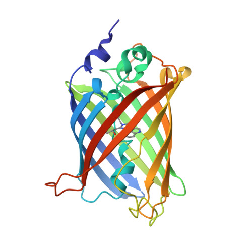 | |
UniProt | |||||
Find proteins for P42212 (Aequorea victoria) Explore P42212 Go to UniProtKB: P42212 | |||||
Entity Groups | |||||
| Sequence Clusters | 30% Identity50% Identity70% Identity90% Identity95% Identity100% Identity | ||||
| UniProt Group | P42212 | ||||
Sequence AnnotationsExpand | |||||
| |||||
| Ligands 2 Unique | |||||
|---|---|---|---|---|---|
| ID | Chains | Name / Formula / InChI Key | 2D Diagram | 3D Interactions | |
| SO4 Query on SO4 | B [auth A] | SULFATE ION O4 S QAOWNCQODCNURD-UHFFFAOYSA-L |  | ||
| ACT Query on ACT | C [auth A] | ACETATE ION C2 H3 O2 QTBSBXVTEAMEQO-UHFFFAOYSA-M |  | ||
| Modified Residues 1 Unique | |||||
|---|---|---|---|---|---|
| ID | Chains | Type | Formula | 2D Diagram | Parent |
| CRF Query on CRF | A | L-PEPTIDE LINKING | C17 H18 N4 O4 |  | THR, TRP, GLY |
| Length ( Å ) | Angle ( ˚ ) |
|---|---|
| a = 51.06 | α = 90 |
| b = 62.64 | β = 90 |
| c = 70.95 | γ = 90 |
| Software Name | Purpose |
|---|---|
| HKL-2000 | data collection |
| PHASES | phasing |
| REFMAC | refinement |
| HKL-2000 | data reduction |
| HKL-2000 | data scaling |
RCSB PDB (citation) is hosted by
RCSB PDB is a member of the

