4HEX

A novel conformation of calmodulin


Experimental Data Snapshot

  • Method: X-RAY DIFFRACTION
  • Resolution: 2.00 Å
  • R-Value Free: 0.237 
  • R-Value Work: 0.210 
  • R-Value Observed: 0.211 

wwPDB Validation   3D Report Full Report


This is version 1.2 of the entry. See complete history


Literature

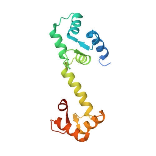
A novel trans conformation of ligand-free calmodulin

Kumar, V.Chichili, V.P.R.Tang, X.Sivaraman, J.

(2013) PLoS One 8: e54834-e54834

  • DOI: https://doi.org/10.1371/journal.pone.0054834
  • Primary Citation of Related Structures:  
    4HEX

  • PubMed Abstract: 

    Calmodulin (CaM) is a highly conserved eukaryotic protein that binds specifically to more than 100 target proteins in response to calcium (Ca(2+)) signal. CaM adopts a considerable degree of structural plasticity to accomplish this physiological role; however, the nature and extent of this plasticity remain to be fully understood. Here, we report the crystal structure of a novel trans conformation of ligand-free CaM where the relative disposition of two lobes of CaM is different, a conformation to-date not reported. While no major structural changes were observed in the independent N- and C-lobes as compared with previously reported structures of Ca(2+)/CaM, the central helix was tilted by ~90° at Arg75. This is the first crystal structure of CaM to show a drastic conformational change in the central helix, and reveals one of several possible conformations of CaM to engage with its binding partner.


  • Organizational Affiliation

    Department of Biological Sciences, National University of Singapore, Republic of Singapore, Republic of Singapore.


Macromolecules
Find similar proteins by:  (by identity cutoff)  |  3D Structure
Entity ID: 1
MoleculeChains Sequence LengthOrganismDetailsImage
Calmodulin
A, B
156Mus musculusMutation(s): 0 
Gene Names: Cam
UniProt
Find proteins for P0DP26 (Mus musculus)
Explore P0DP26 
Go to UniProtKB:  P0DP26
Entity Groups  
Sequence Clusters30% Identity50% Identity70% Identity90% Identity95% Identity100% Identity
UniProt GroupP0DP26
Sequence Annotations
Expand
  • Reference Sequence
Small Molecules
Ligands 2 Unique
IDChains Name / Formula / InChI Key2D Diagram3D Interactions
ZN
Query on ZN

Download Ideal Coordinates CCD File 
C [auth A]
E [auth A]
G [auth A]
H [auth B]
K [auth B]
C [auth A],
E [auth A],
G [auth A],
H [auth B],
K [auth B],
L [auth B]
ZINC ION
Zn
PTFCDOFLOPIGGS-UHFFFAOYSA-N
CA
Query on CA

Download Ideal Coordinates CCD File 
D [auth A],
F [auth A],
I [auth B],
J [auth B]
CALCIUM ION
Ca
BHPQYMZQTOCNFJ-UHFFFAOYSA-N
Experimental Data & Validation

Experimental Data

  • Method: X-RAY DIFFRACTION
  • Resolution: 2.00 Å
  • R-Value Free: 0.237 
  • R-Value Work: 0.210 
  • R-Value Observed: 0.211 
  • Space Group: P 1 21 1
Unit Cell:
Length ( Å )Angle ( ˚ )
a = 38.827α = 90
b = 116.397β = 94.83
c = 38.797γ = 90
Software Package:
Software NamePurpose
HKL-2000data collection
SHELXCDphasing
SHELXEmodel building
PHENIXrefinement
HKL-2000data reduction
HKL-2000data scaling
REFMACrefinement

Structure Validation

View Full Validation Report


Entry History 

Deposition Data

Revision History  (Full details and data files)

  • Version 1.0: 2013-03-06
    Type: Initial release
  • Version 1.1: 2017-11-15
    Changes: Refinement description
  • Version 1.2: 2024-03-20
    Changes: Data collection, Database references, Derived calculations, Refinement description