4IOF

Crystal structure analysis of Fab-bound human Insulin Degrading Enzyme (IDE)


Experimental Data Snapshot

  • Method: X-RAY DIFFRACTION
  • Resolution: 3.35 Å
  • R-Value Free: 0.280 
  • R-Value Work: 0.232 
  • R-Value Observed: 0.235 

wwPDB Validation   3D Report Full Report


This is version 1.4 of the entry. See complete history


Literature

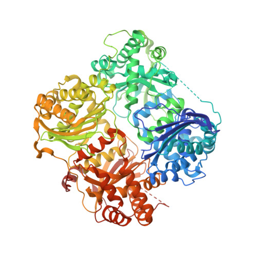
Conformational states and recognition of amyloidogenic peptides of human insulin-degrading enzyme.

McCord, L.A.Liang, W.G.Dowdell, E.Kalas, V.Hoey, R.J.Koide, A.Koide, S.Tang, W.J.

(2013) Proc Natl Acad Sci U S A 110: 13827-13832

  • DOI: https://doi.org/10.1073/pnas.1304575110
  • Primary Citation of Related Structures:  
    4IOF

  • PubMed Abstract: 

    Insulin-degrading enzyme (IDE) selectively degrades the monomer of amyloidogenic peptides and contributes to clearance of amyloid β (Aβ). Thus, IDE retards the progression of Alzheimer's disease. IDE possesses an enclosed catalytic chamber that engulfs and degrades its peptide substrates; however, the molecular mechanism of IDE function, including substrate access to the chamber and recognition, remains elusive. Here, we captured a unique IDE conformation by using a synthetic antibody fragment as a crystallization chaperone. An unexpected displacement of a door subdomain creates an ~18-Å opening to the chamber. This swinging-door mechanism permits the entry of short peptides into the catalytic chamber and disrupts the catalytic site within IDE door subdomain. Given the propensity of amyloidogenic peptides to convert into β-strands for their polymerization into amyloid fibrils, they also use such β-strands to stabilize the disrupted catalytic site resided at IDE door subdomain for their degradation by IDE. Thus, action of the swinging door allows IDE to recognize amyloidogenicity by substrate-induced stabilization of the IDE catalytic cleft. Small angle X-ray scattering (SAXS) analysis revealed that IDE exists as a mixture of closed and open states. These open states, which are distinct from the swinging door state, permit entry of larger substrates (e.g., Aβ, insulin) to the chamber and are preferred in solution. Mutational studies confirmed the critical roles of the door subdomain and hinge loop joining the N- and C-terminal halves of IDE for catalysis. Together, our data provide insights into the conformational changes of IDE that govern the selective destruction of amyloidogenic peptides.


  • Organizational Affiliation

    Ben-May Department for Cancer Research, University of Chicago, Chicago, IL 60637, USA.


Macromolecules
Find similar proteins by:  (by identity cutoff)  |  3D Structure
Entity ID: 1
MoleculeChains Sequence LengthOrganismDetailsImage
Insulin-degrading enzyme
A, B
990Homo sapiensMutation(s): 0 
Gene Names: IDEInsulin Degrading Enzyme
EC: 3.4.24.56
UniProt & NIH Common Fund Data Resources
Find proteins for P14735 (Homo sapiens)
Explore P14735 
Go to UniProtKB:  P14735
PHAROS:  P14735
GTEx:  ENSG00000119912 
Entity Groups  
Sequence Clusters30% Identity50% Identity70% Identity90% Identity95% Identity100% Identity
UniProt GroupP14735
Sequence Annotations
Expand
  • Reference Sequence
Find similar proteins by:  (by identity cutoff)  |  3D Structure
Entity ID: 2
MoleculeChains Sequence LengthOrganismDetailsImage
Fab-bound IDE, heavy chain
C, E
263Mus musculusMutation(s): 0 
Entity Groups  
Sequence Clusters30% Identity50% Identity70% Identity90% Identity95% Identity100% Identity
Sequence Annotations
Expand
  • Reference Sequence
Find similar proteins by:  (by identity cutoff)  |  3D Structure
Entity ID: 3
MoleculeChains Sequence LengthOrganismDetailsImage
Fab-bound IDE, light chain
D, F
239Mus musculusMutation(s): 0 
Entity Groups  
Sequence Clusters30% Identity50% Identity70% Identity90% Identity95% Identity100% Identity
Sequence Annotations
Expand
  • Reference Sequence
Small Molecules
Ligands 1 Unique
IDChains Name / Formula / InChI Key2D Diagram3D Interactions
ZN
Query on ZN

Download Ideal Coordinates CCD File 
G [auth A]ZINC ION
Zn
PTFCDOFLOPIGGS-UHFFFAOYSA-N
Experimental Data & Validation

Experimental Data

  • Method: X-RAY DIFFRACTION
  • Resolution: 3.35 Å
  • R-Value Free: 0.280 
  • R-Value Work: 0.232 
  • R-Value Observed: 0.235 
  • Space Group: P 21 21 21
Unit Cell:
Length ( Å )Angle ( ˚ )
a = 56.955α = 90
b = 131.662β = 90
c = 377.039γ = 90
Software Package:
Software NamePurpose
HKL-3000data collection
PHASESphasing
PHENIXrefinement
HKL-3000data reduction
HKL-3000data scaling

Structure Validation

View Full Validation Report


Entry History 

Deposition Data

Revision History  (Full details and data files)

  • Version 1.0: 2013-07-24
    Type: Initial release
  • Version 1.1: 2013-08-21
    Changes: Database references
  • Version 1.2: 2013-09-04
    Changes: Database references
  • Version 1.3: 2018-01-24
    Changes: Structure summary
  • Version 1.4: 2023-09-20
    Changes: Data collection, Database references, Derived calculations, Refinement description