


Co-crystallization of streptavidin-biotin complex with a lanthanide-ligand complex gives rise to a novel crystal form
Bandara, R.A.M.S.S., Liu, D.Q., Hindupur, A., Tesh, K.F., Fox, R.O.To be published.
Experimental Data Snapshot
Entity ID: 1 | |||||
|---|---|---|---|---|---|
| Molecule | Chains | Sequence Length | Organism | Details | Image |
| Streptavidin | 129 | Streptomyces avidinii | Mutation(s): 0 | 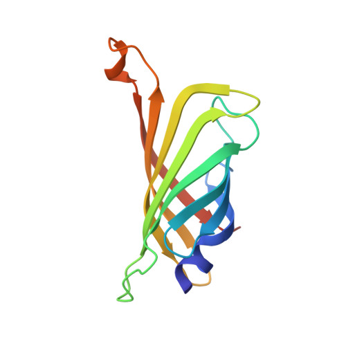 | |
UniProt | |||||
Find proteins for P22629 (Streptomyces avidinii) Explore P22629 Go to UniProtKB: P22629 | |||||
Entity Groups | |||||
| Sequence Clusters | 30% Identity50% Identity70% Identity90% Identity95% Identity100% Identity | ||||
| UniProt Group | P22629 | ||||
Sequence AnnotationsExpand | |||||
| |||||
| Ligands 4 Unique | |||||
|---|---|---|---|---|---|
| ID | Chains | Name / Formula / InChI Key | 2D Diagram | 3D Interactions | |
| BTN Query on BTN | N [auth A] | BIOTIN C10 H16 N2 O3 S YBJHBAHKTGYVGT-ZKWXMUAHSA-N |  | ||
| PDC Query on PDC | B [auth A] C [auth A] D [auth A] E [auth A] F [auth A] | PYRIDINE-2,6-DICARBOXYLIC ACID C7 H5 N O4 WJJMNDUMQPNECX-UHFFFAOYSA-N |  | ||
| TB Query on TB | O [auth A], P [auth A], Q [auth A], R [auth A], S [auth A] | TERBIUM(III) ION Tb HKCRVXUAKWXBLE-UHFFFAOYSA-N |  | ||
| NA Query on NA | T [auth A] | SODIUM ION Na FKNQFGJONOIPTF-UHFFFAOYSA-N |  | ||
| Length ( Å ) | Angle ( ˚ ) |
|---|---|
| a = 64.423 | α = 90 |
| b = 71.395 | β = 90 |
| c = 55.494 | γ = 90 |
| Software Name | Purpose |
|---|---|
| d*TREK | data scaling |
| d*TREK | data reduction |
| PHENIX | refinement |
| PDB_EXTRACT | data extraction |
| PHENIX | phasing |
RCSB PDB (citation) is hosted by
RCSB PDB is a member of the

